Created 2008.
Acquired benign keratinocytic tumours to be discussed in this section include seborrhoeic keratoses, corns and calluses. They are extremely common and mainly of cosmetic concern. They may occasionally prove painful.
There are various benign tumours derived from adnexal structures. Various combinations of follicular, sebaceous, apocrine and eccrine differentiation occur commonly (mixed tumour or chondroid syringoma).
Seborrhoeic keratoses (basal cell papillomas) appear as stuck-on warty plaques and are very common on the face, neck and trunk in those over 40 years of age. The surface may be waxy or rough. They do not affect non-hair bearing areas such as the palms and soles. There is a familial predisposition in those with hundreds of lesions. They can present as macules, papules, plaques, verrucous, polypoid or pedunculated lesions. They can range in colour from white to black and may be confused with malignant melanoma; dermatoscopic features are often helpful to enable the distinction.
Some solar lentigines represent flat seborrhoeic warts.
Multiple eruptive lesions may arise when a patient is unwell, and is sometimes a sign of systemic malignancy (sign of Leser-Trélat. They may also appear after skin injury, including sun burn and dermatitis.
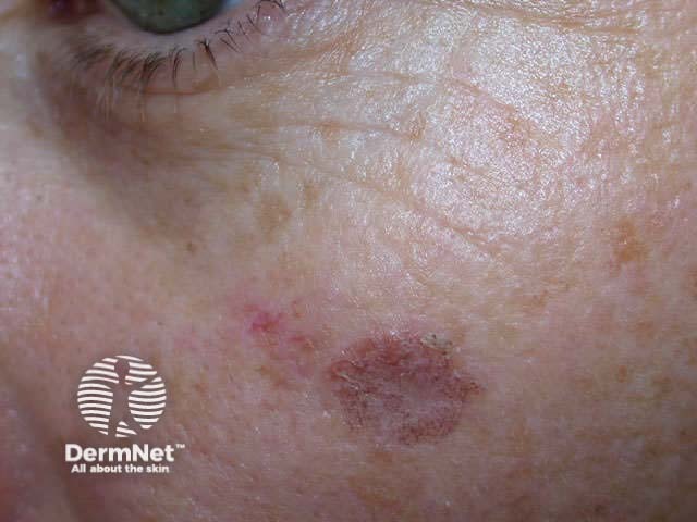
Irritated seborrhoeic keratosis is erythematous and crusty. It can resemble squamous cell carcinoma. These lesions may resolve spontaneously.


Variants include:


Seborrhoeic keratoses are unsightly and often pruritic. They are readily removed by superficial destructive means such as shave excision, electrodessication or cryotherapy. However, if melanoma is considered in the differential diagnosis, the lesions should be fully excised for histology.
Corns and calluses represent localised hyperkeratosis induced by pressure, most often on hands and feet. Hard corns are firm papules with translucent central cores, found on the dorsolateral fifth toes and dorsal aspects of other toes. Soft corns are painful scaly plaques found between the toes. They often become macerated. Broad keratotic plaques, often under the metatarsal heads or over the metacarpals, are calluses.


Treatment involves paring down or filing the callosity. This is easier after soaking in warm water or applying a keratolytic such as salicylic acid or urea ointment, gel, paint or plaster. The pressure on the affected area should be relieved where possible using soft cushioning material or specific orthotic devices.
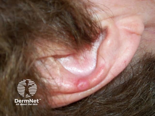
This condition, often referred to as ‘CNH’, presents as a tender papule on one or both ears, most often affecting the helix or antehelix. It is most common in men over 40 years, and probably represents ischaemic damage due to pressure on sun- and/or cold-damaged skin and cartilage, provoked by lying on it at night. Treatment involves relieving the pressure using a foam or silicone protector. Persistent lesions are usually excised but may recur.


There are many types of cutaneous cyst.
Epidermoid cysts are walled-off cavities filled with keratin and are derived from the hair follicle unit. Their contents can be expressed and have an unpleasant, cheesy odour. Epidermoid cysts are often found on the face and upper trunk and may be an inherited trait. If they rupture, they become inflamed (red and painful). Although this resembles infection, culture is generally sterile and antibiotics ineffective.


Small superficial and often multiple cysts are known as milia, and occur on the face or as inclusion cysts in healing wounds, characteristically after subepidermal blistering.


Multiple epidermoid cysts may accompany acne vulgaris. They are also a feature of Gardner syndrome, when they are associated with colonic polyps. Multiple scrotal or vulval cysts eventually calcify.


Trichilemmal or pilar cysts (scalp), vellus hair cysts (eruptive papules) and steatocystoma (oily lesions on chest) have characteristic histological features. The dermoid cyst occurs at the site of embryonic fusion planes, for example lateral eyebrow. A preauricular cyst is often called an ‘ear pit cyst’. The pilonidal cyst arises in the upper gluteal cleft, and may be associated with hidradenitis suppurativa.


Cysts composed of non-stratified epithelium include hidrocystoma (eyelid margin) and others described by their embryonic origin, such as bronchogenic cyst (suprasternal notch), thyroglossal duct cyst (midline anterior neck), brachial cleft cyst (lateral neck and mandible).
Non-epithelial cysts include the mucocoele (inner lip), mucous cyst (distal digit and nail bed), ganglion (wrist) pseudocysts of auricle (ear) and some traumatic cysts.


Lesions showing follicular differentiation contain basaloid cells resembling the follicular bulb, adjacent mesenchymal cells resembling the papilla, ‘shadow’ cells and trichilemmal (outer root sheath) cells, i.e. palisading pale cells with a thickened basement membrane.
Benign solitary or multiple follicular tumours include:


Sebaceous differentiation results in cells with coarsely vacuolated cytoplasm and scalloped nuclei.
Sebaceous hyperplasia represents benign enlargement of sebaceous lobules around the follicular infundibulum. Yellowish papules appear on the forehead or cheeks and have a characteristic central dimple.


These are solitary or multiple yellowish papules or nodules in which there is a proliferation of sebocytes and/or seboblasts (Muir-Torre syndrome)
It is difficult to determine whether sweat gland tumours are of eccrine or apocrine differentiation. The precise diagnosis of sweat gland lesions is most likely to be made histologically rather than clinically.
Syringomata are sweat duct tumours most often presenting as multiple skin-coloured papules on the eyelids. They may also develop on genital skin (apocrine). Eruptive syringomata sometimes affect hands and feet (eccrine).


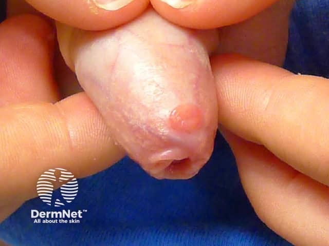
Poromas have terminal ductal differentiation. They present as solitary papules most often on acral regions (eccrine origin) and are sometimes pigmented. Hidradenoma looks similar, but the cells have a greater amount of cytoplasm histologically. They most often appear in genital sites (apocrine origin). Papillary hidradenoma (hidradenoma papilliferum) is a variant with papillary pattern histologically, and may affect the nipple. Syringocystadenoma papilliferum tends to present as grouped keratotic papules, and may arise in a sebaceous naevus so presumably also has apocrine origin.


Spiradenoma is the name given to an undifferentiated or poorly differentiated benign adnexal neoplasm that has some tubular features. They may be solitary or multiple and are often painful.
Cylindroma describes an undifferentiated or poorly differentiated adnexal neoplasm with a mosaic pattern, presumably apocrine in origin. They may be solitary or multiple and are sometimes huge (turban tumour).
Paget disease presents as a dry red mildly irritable plaque. It is due to an intraepidermal malignant proliferation of clear cells and has characteristic histology.
Mammary Paget disease presents as a unilateral eczema-like plaque on the nipple or areola. It is associated with underlying in-situ ductal carcinoma or other underlying cancer. Investigation should include mammography and management should be by a breast surgeon.
Extra-mammary Paget disease presents as a dry red plaque in the axillae, or on genital or perianal skin. It is sometimes associated with underlying adenocarcinoma arising in adjacent but not necessarily contiguous tissue. If no such tumour is found, wide local excision is the usual management but recurrence is common. Alternative treatment may include fluorouracil cream, imiquimod cream, or photodynamic therapy.


Describe Birt-Hogg-Dubé syndrome.
Information for patients
See the DermNet bookstore

