Created 2009.
The dermatoses described on this page are the result of an immunological reaction to underlying systemic disease. In each case, a number of different conditions may be responsible. The severity of the reaction does not necessarily correllate with the severity of the underlying disease.
The mechanisms for the reactions are poorly understood.
Panniculitis is the term used for a group of disorders in which inflammation primarily affects subcutaneous tissue.
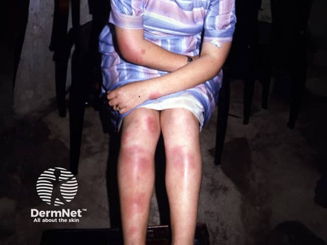
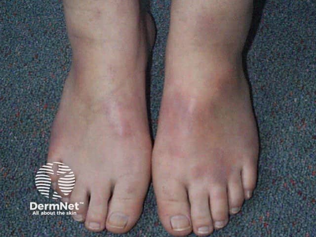
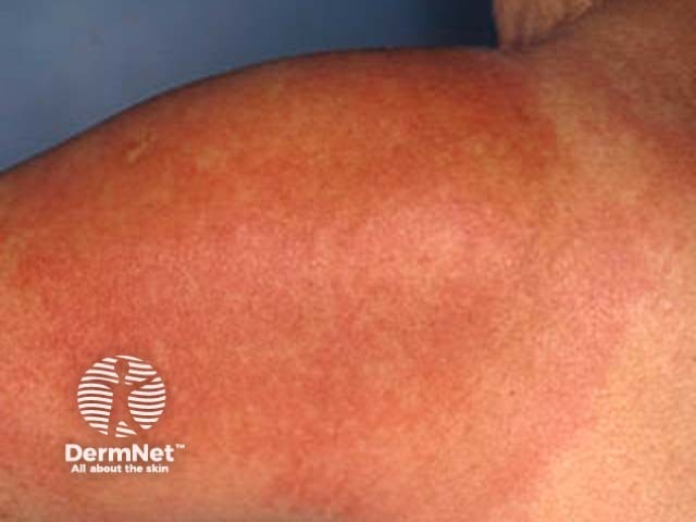
Erythema nodosum
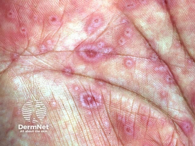
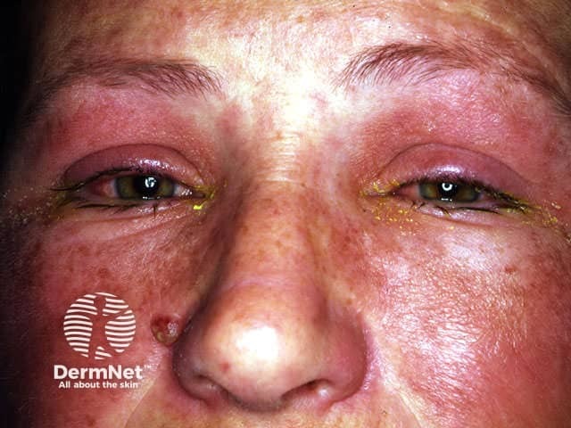
Erythema multiforme (EM) is an acute eruption of erythematous target or iris-shaped plaques that may blister, typically involving the extremities (face, palms, soles and distal limbs).
EM minor
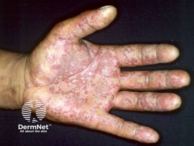
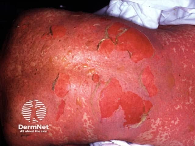
EM major
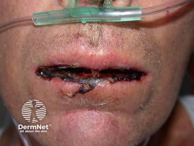
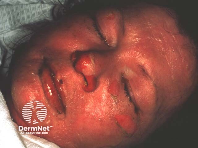
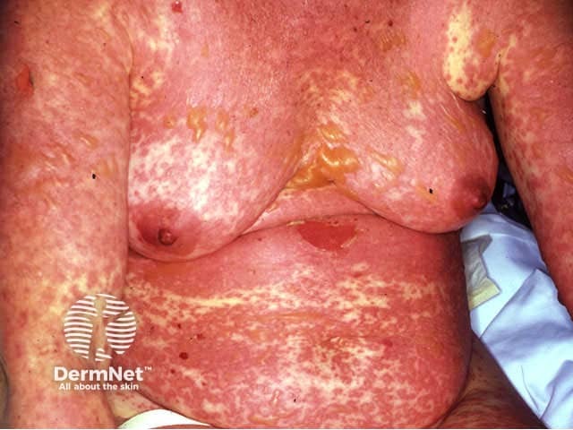
Toxic epidermal necrolysis
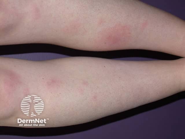
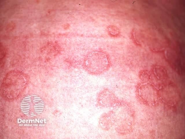
Several patterns of annular erythema are described, sometimes specific to the underlying aetiology.
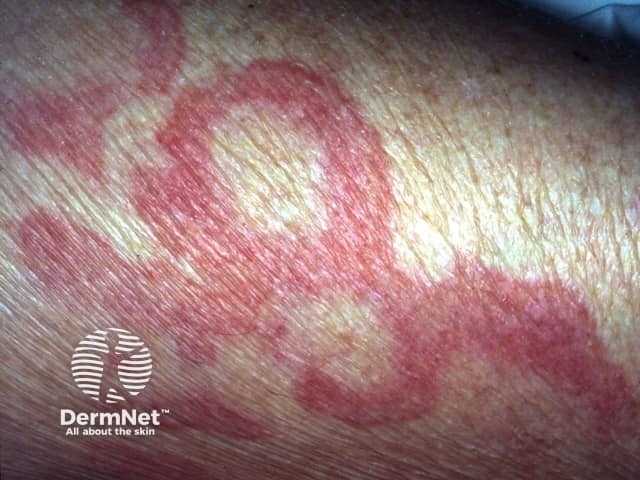
Erythema annulare centrifugum results in expanding erythematous annular and polycyclic plaques, generally on the trunk.
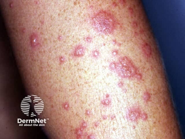
Dermatoses with dense dermal infiltrates of neutrophils include:
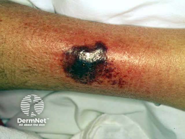
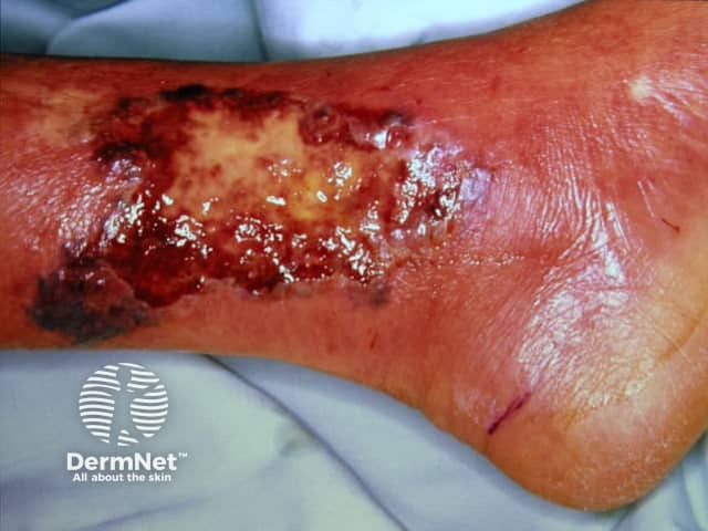
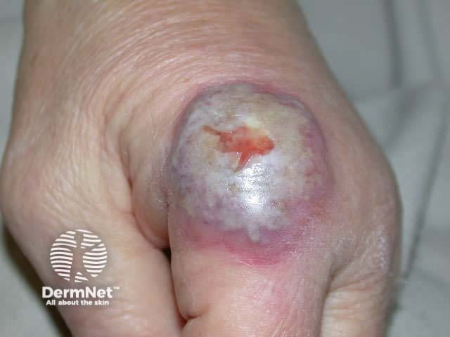
Pyoderma gangrenosum is associated with inflammatory bowel disease, rheumatoid arthritis and haematological malignancies but occasionally arises in otherwise healthy individuals. Sweet disease can have similar causes but more often follows an upper respiratory tract infection.
Pyoderma gangrenosum results in severe ulceration characterised by an overhanging necrotic edge and severe pain. It most commonly arises on the lower legs and may be very difficult to control requiring one or more systemic immune suppressive medications.
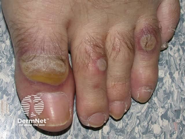
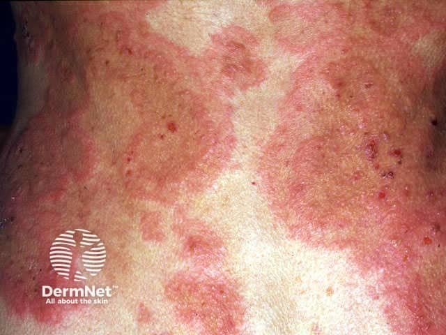
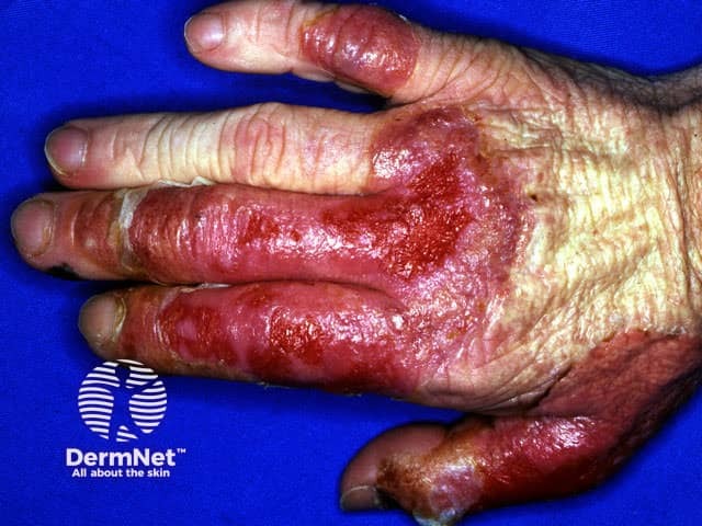
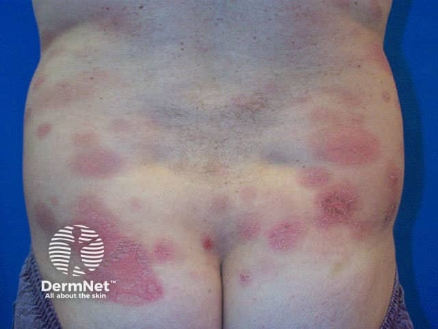
Sweet disease presents as rapidly evolving juicy “pseudo-vesicular” plaques accompanied by fever, leukocytosis, conjunctivitis and arthralgia. The lesions are most often located asymmetrically on the upper extremities, neck and face (cape distribution). They heal within two or three months without scarring, but resolve very quickly if treated with oral prednisone for 4 to 6 weeks.
What is the evidence that systemic corticosteroids should be avoided in the management of Stevens Johnson syndrome and toxic epidermal necrolysis?
Information for patients
See the DermNet bookstore

