DermNet provides Google Translate, a free machine translation service. Note that this may not provide an exact translation in all languages

Translate

Cutaneous lupus erythematosus

Author: Amanda Oakley, Dermatologist, Hamilton, New Zealand, 2001/2016

DermNet Update April 2021; further minor update October 2023.


toc-icon

What is cutaneous lupus erythematosus?

Cutaneous lupus erythematosus (LE) is a diverse group of autoimmune connective tissue disorders localised to the skin that can be associated with systemic lupus erythematosus (SLE) to varying degrees.

Cutaneous lupus erythematosus (CLE) is classified as:

Cutaneous lupus erythematosus

Skin lesions of cutaneous lupus erythematosus can be subdivided into:

  • Lupus-specific changes
    • Clinical signs diagnostic of, or unique to, LE
    • Histology is diagnostic for LE; vacuolar interface dermatitis
  • Lupus-nonspecific changes
    • Clinical signs found not only in LE, but also in other connective tissue diseases
    • Histology is not diagnostic for LE; reactive patterns seen.

Who gets cutaneous lupus erythematosus?

Cutaneous lupus erythematosus has an annual incidence of 4 cases per 100,000 people, and a prevalence of 73 cases per 100,000.

As with SLE, there is a marked female predominance with CLE particularly affecting women 20 to 50 years of age. However, all age groups and both sexes can be affected. Skin of colour is an important predisposing factor.

Up to 75% of patients with SLE develop cutaneous signs, and these may be the first indication of SLE in 25%.

What causes cutaneous lupus erythematosus?

The pathogenesis of cutaneous lupus erythematosus is multifactorial:

  • Genetic susceptibility
    • High incidence among family members
  • Environmental factors
    • Cigarette smoking
    • Sun exposure
    • Medications
  • Innate and adaptive immune responses
    • autoantibodies.

What are the clinical features of cutaneous lupus erythematosus?

This is a brief overview of the clinical features of cutaneous lupus erythematosus. See the specific lupus pages for more detail – links are provided at the end under the ‘On DermNet’ heading.

Acute cutaneous lupus erythematosus

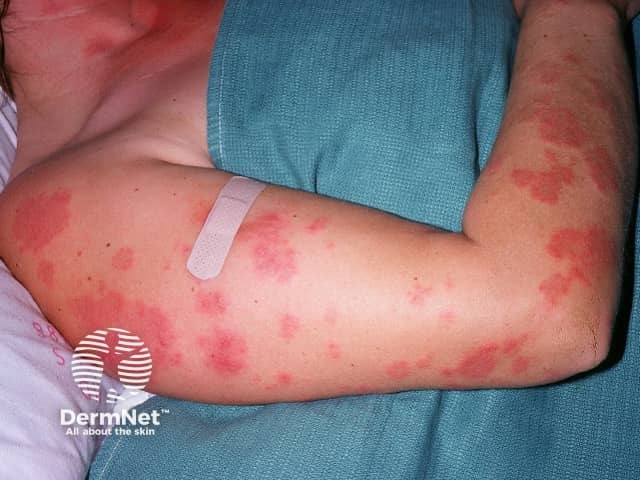
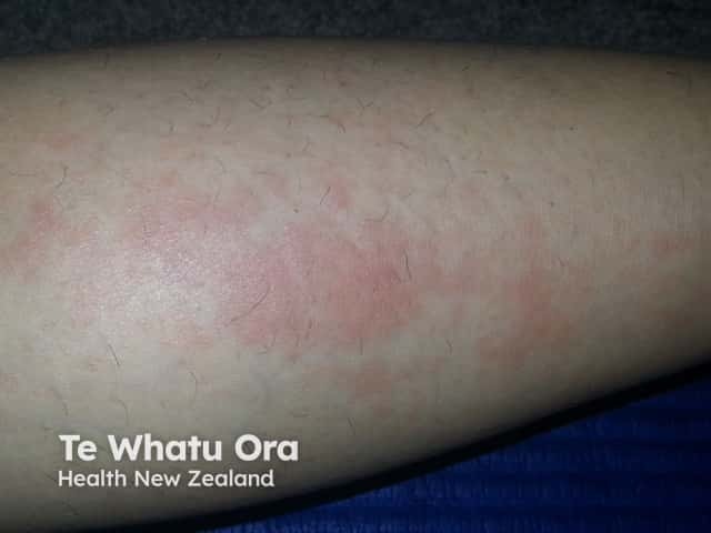
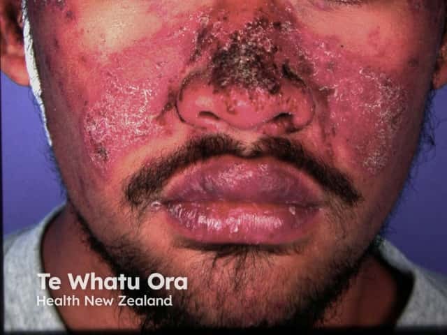
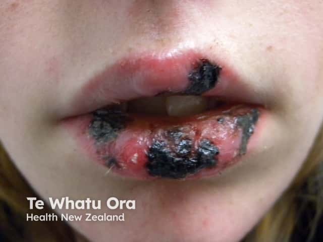
Acute cutaneous lupus erythematosus typically presents as transient erythematous patches associated with a flare of systemic lupus erythematosus.

Lupus-specific skin changes:

  • Localised acute CLE: malar ‘butterfly' rash – redness and swelling over both cheeks, sparing the nasolabial folds, lasting hours to days
  • Generalised acute CLE: diffuse or papular erythema of the face, upper limbs (sparing the knuckles), and trunk resembling a morbilliform drug eruption or viral exanthem
  • Toxic epidermal necrolysis-like acute CLE: is associated with lupus nephritis or cerebritis, and must be distinguished from drug-induced toxic epidermal necrolysis in a patient with SLE.

Acute cutaneous lupus erythematosus

Subacute cutaneous lupus erythematosus

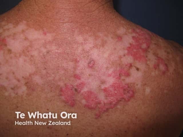
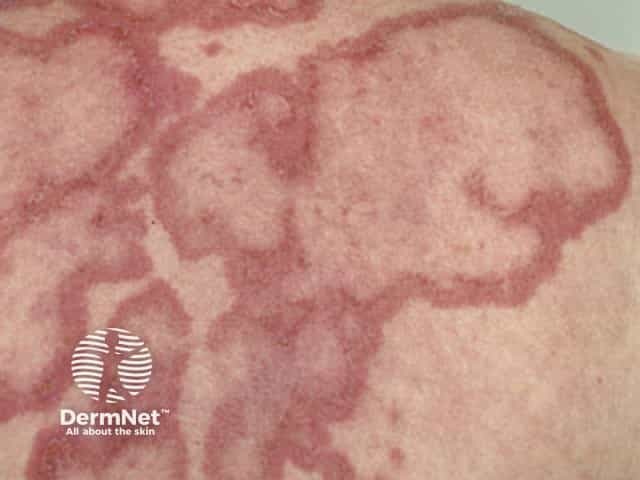
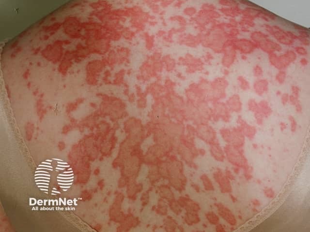
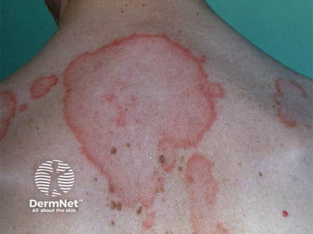
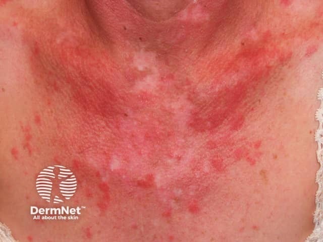
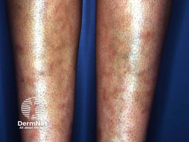
Subacute cutaneous lupus erythematosus is less commonly associated with SLE with approximately 50% having a mild form of SLE. It is thought 20–40% have drug-induced SCLE. It comprises 10–15% of cutaneous LE presentations. The skin changes are more persistent than those of ACLE.

Skin lesions of SCLE:

  • Occur on the trunk and upper limbs, triggered or aggravated by sun exposure
  • Present as a psoriasiform papulosquamous rash or annular, polycyclic plaques with central clearing
  • Resolve to leave dyspigmentation and telangiectases, but no scarring.

Subacute cutaneous lupus erythematosus

Intermittent cutaneous lupus erythematosus

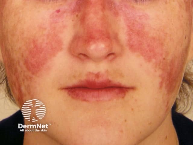
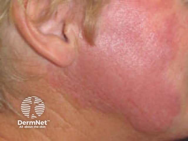
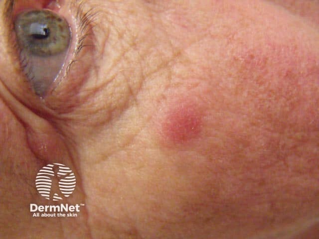
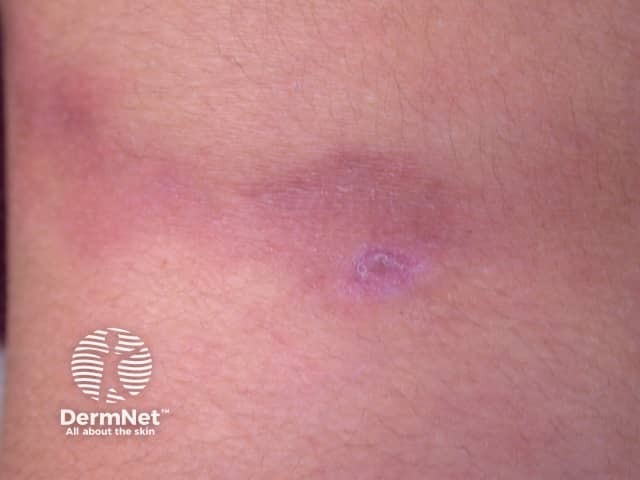
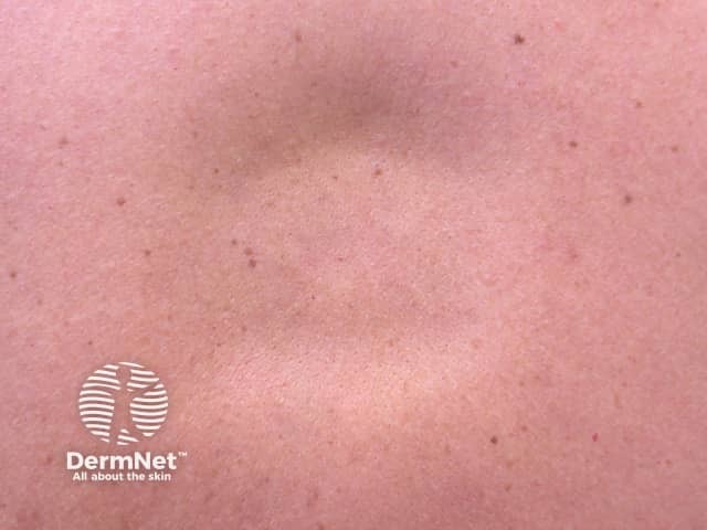
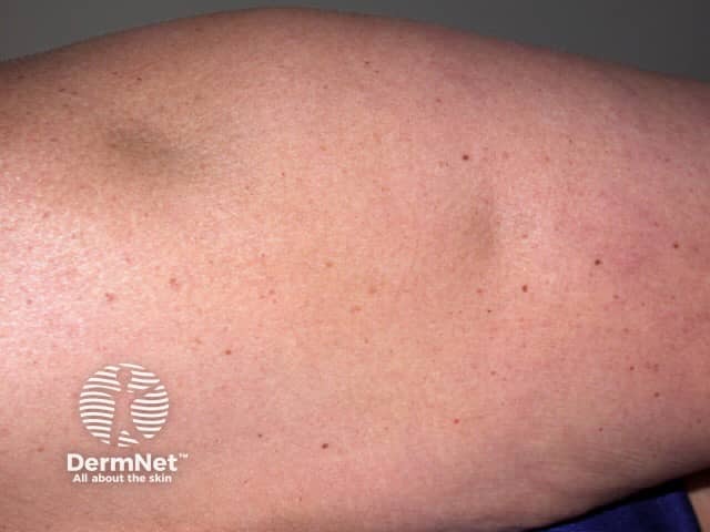
Intermittent CLE is better known as lupus tumidus, a dermal form of lupus erythematosus.

Skin lesions of lupus tumidus:

  • Occur on sun-exposed areas of skin, such as the face, neck, and upper anterior chest
  • Present as erythematous, round or annular, papules and plaques with a smooth surface
  • Resolve in winter without scarring.

Lupus tumidus

Chronic cutaneous lupus erythematosus

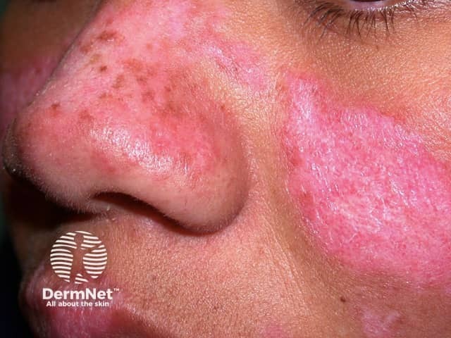
Chronic cutaneous lupus erythematosus is the most common form of CLE, and about 25% of SLE patients have some form of CCLE.

Discoid lupus erythematosus

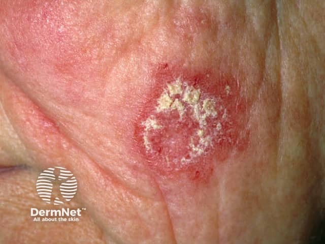
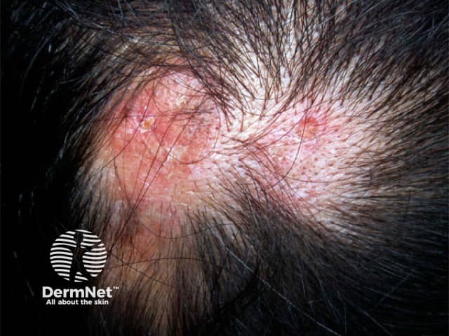
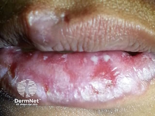
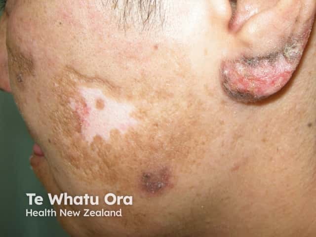
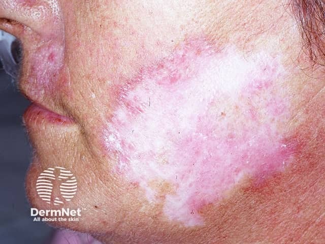
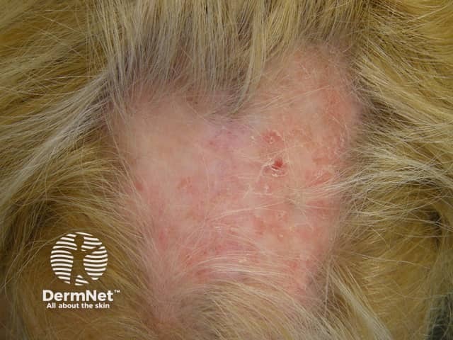
Discoid lupus erythematosus is the most common form of CCLE (80%) and is particularly prevalent and severe in patients with skin of colour. Only 1–2% of patients with localised DLE progress to SLE.

Skin lesions of DLE:

  • Are most commonly located on the scalp, ears, cheeks, nose, and lips
  • Present as destructive scaly plaques with follicular prominence (carpet tack sign) which can result in scarring alopecia
  • Heal slowly leaving post-inflammatory dyspigmentation and scarring.

Discoid lupus erythematosus

Lupus profundus

Lupus profundus is a mostly lobular panniculitis without vasculitis.

Skin lesions of lupus profundus:

  • Can develop at any site
  • Present as persistent, firm, deep, tender nodules
  • Resolve to leave dents in the skin due to atrophy of the fat.

Lupus profundus

Chilblain lupus erythematosus

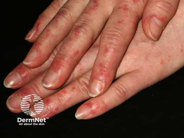
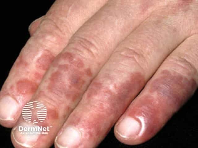
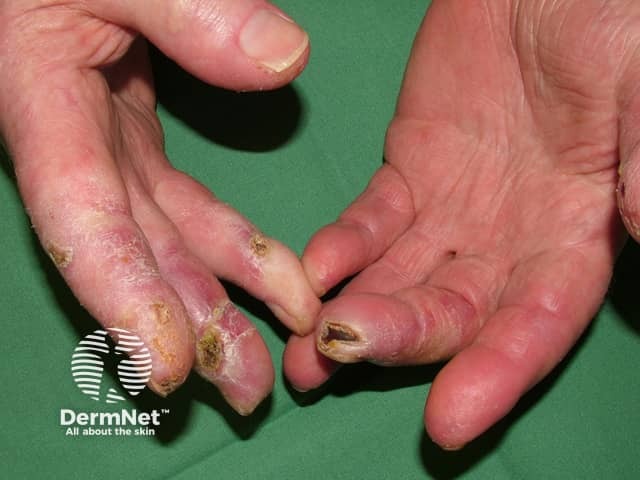
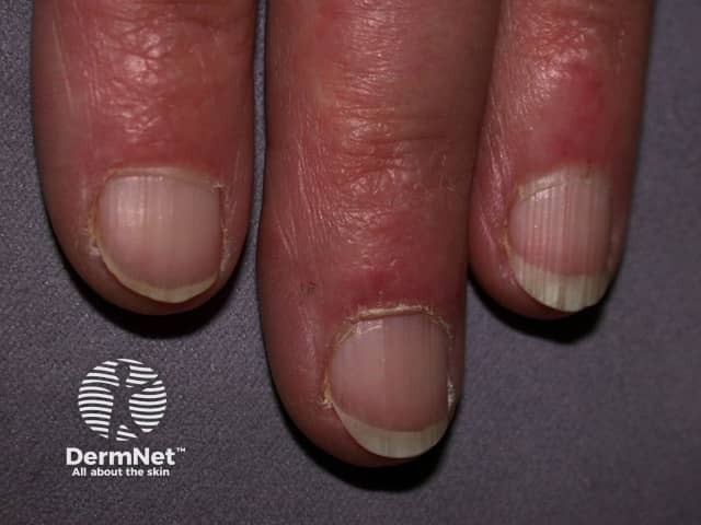
Chilblain lupus erythematosus is an under-reported form of chronic CLE involving mainly the fingers and toes of smokers triggered by exposure to a moist cold environment. It may be familial with no association to SLE, or sporadic which can be associated with SLE.

Skin lesions of chilblain lupus:

  • Are often pruritic and painful
  • May ulcerate or develop hyperkeratotic fissuring
  • May heal leaving depigmentation and atrophic spindling of the distal phalange.

Chilblain lupus erythematosus

How do clinical features vary in differing types of skin?

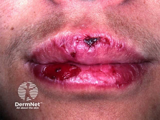
Some forms of cutaneous lupus erythematosus, such as bullous lupus erythematosus and discoid lupus erythematosus, are particularly associated with skin of colour.

Dyspigmentation and scarring are complications of CLE that significantly impact quality of life for patients with skin of colour.

Cutaneous lupus erythematosus in skin of colour

Non-specific cutaneous lupus erythematosus

Non-specific skin signs of systemic lupus erythematosus

What are the complications of cutaneous lupus erythematosus?

Complications of cutaneous lupus erythematosus

How is cutaneous lupus erythematosus diagnosed?

  • Skin biopsy — diagnostic histopathology and direct immunofluorescence is seen only in specific-LE lesions [see Discoid lupus erythematosus pathology]
  • Blood tests — full blood count, renal function test, inflammatory markers
  • Serology — including ANA, ENA – are often negative in chronic CLE

Cutaneous Lupus Erythematosus Disease Area and Severity Index (CLASI)

The Cutaneous Lupus Erythematosus Disease Area and Severity Index scores activity and damage in each of 12 anatomical locations.

The total activity score:

  • Degree of redness (0-3) and scale (0-2)
  • Mucous membrane involvement (0-1)
  • Recent hair loss (0-1), nonscarring alopecia (0-3)

Total damage score:

  • Degree of dyspigmentation (0-2) and scarring (0-2)
  • Persistence of dyspigmentation more than 12 months doubles the dyspigmentation score
  • Scalp scarring (0, 3, 4, 5, 6).

What is the treatment for cutaneous lupus erythematosus?

General measures

Specific measures

Local therapy

Systemic therapy

What is the outcome for cutaneous lupus erythematosus?

Cutaneous lupus erythematosus can be the presenting sign of SLE, as in acute CLE, or may evolve into SLE.

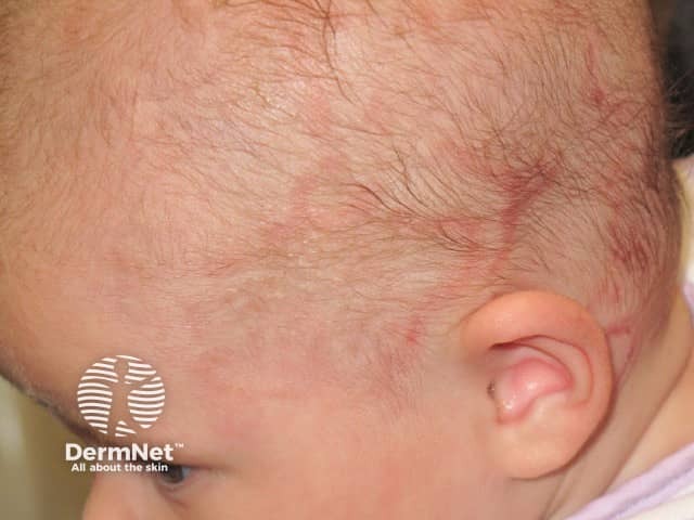
Female patients with CLE and Ro/La autoantibodies should be advised of the risk their baby may have neonatal lupus erythematosus including congenital heart block.

Chronic CLE tends to follow a chronic relapsing course for years, with flares in spring and summer, and resolution with scarring if untreated.


References

  • Chakka S, Krain RL, Concha JSS, Chong BF, Merola JF, Werth VP. The CLASI, a validated tool for the evaluation of skin disease in lupus erythematosus: a narrative review. Ann Transl Med. 2021;9(5):431. doi:10.21037/atm-20-5048. Journal
  • Filotico R, Mastrandrea V. Cutaneous lupus erythematosus: clinico-pathologic correlation. G Ital Dermatol Venereol. 2018;153(2):216–29. doi:10.23736/S0392-0488.18.05929-1. Journal
  • Garelli CJ, Refat MA, Nanaware PP, Ramirez-Ortiz ZG, Rashighi M, Richmond JM. Current insights in cutaneous lupus erythematosus immunopathogenesis. Front Immunol. 2020;11:1353. doi:10.3389/fimmu.2020.01353. Journal
  • Goodfield M, Dutz J, McCourt C. Lupus erythematosus. In: Griffiths C, Barker J, Bleiker T, Chalmers R, Creamer D (eds). Rook’s Textbook of Dermatology, 9th edn, Wiley Blackwell, 2016: 51.1–39.
  • Hersh AO, Arkin LM, Prahalad S. Immunogenetics of cutaneous lupus erythematosus. Curr Opin Pediatr. 2016;28(4):470–5. doi:10.1097/MOP.0000000000000383. PubMed Central
  • Little AJ, Vesely MD. Cutaneous lupus erythematosus: current and future pathogenesis-directed therapies. Yale J Biol Med. 2020;93(1):81–95. PubMed Central
  • Petty AJ, Floyd L, Henderson C, Nicholas MW. Cutaneous lupus erythematosus: progress and challenges. Curr Allergy Asthma Rep. 2020;20(5):12. doi:10.1007/s11882-020-00906-8. PubMed Central
  • Stannard JN, Kahlenberg JM. Cutaneous lupus erythematosus: updates on pathogenesis and associations with systemic lupus. Curr Opin Rheumatol. 2016;28(5):453–9. doi:10.1097/BOR.0000000000000308. PubMed Central

On DermNet

Other websites

Books about skin diseases


Related information

Sign up to the newsletter