DermNet provides Google Translate, a free machine translation service. Note that this may not provide an exact translation in all languages

Translate

Lichenoid keratosis

Author: Vanessa Ngan, Staff Writer, 2006. Updated by A/Prof Amanda Oakley, March 2016. DermNet NZ Revision September 2021


toc-icon

What is lichenoid keratosis?

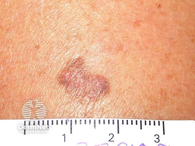
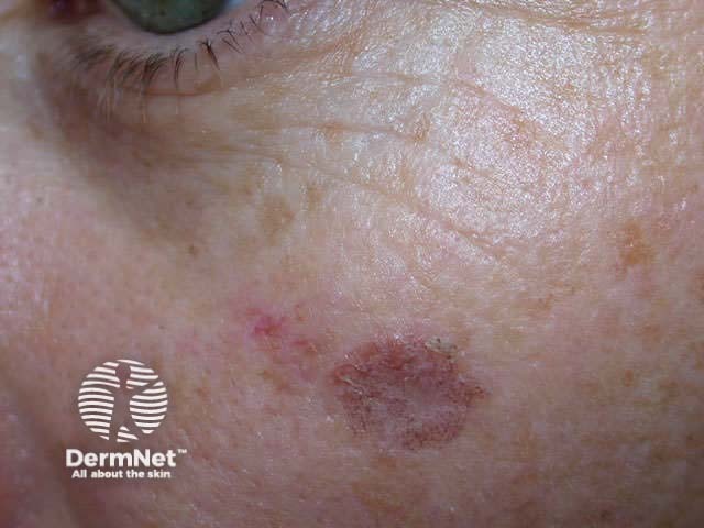
Lichenoid keratosis is a small, inflamed macule or thin pigmented plaque, usually solitary, with a lichenoid tissue reaction on histology.
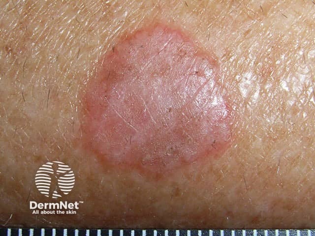
Lichenoid keratosis

Lichenoid keratosis is also known as benign lichenoid keratosis, solitary lichen planus, lichen planus-like keratosis and involuting lichenoid plaque. It is one of the causes of atypical solar lentigo.

Who gets lichenoid keratosis?

Lichenoid keratosis generally develops in fair-skinned patients aged 30–80 years. It is twice as common in females as than males. It is most commonly seen in Caucasians and rarely affects Asians, African Americans, or Hispanics.

What causes lichenoid keratosis?

Lichenoid keratosis is an inflammatory reaction arising in a regressing existing solar lentigo or seborrhoeic keratosis. It is not known what causes the reaction, but triggers can include minor trauma such as friction, drugs, dermatitis, and sun exposure. 

What are the clinical features of lichenoid keratosis?

  • A solitary lesion is present in 90% of cases of lichenoid keratosis, with others presenting with multiple lesions.
  • It is most commonly found on the upper trunk, followed by the distal upper extremities, and less commonly on the head and neck.
  • Size ranges from a few millimetres to one centimetre or more in size.
  • The skin surface may be smooth, scaly, or warty.
  • The lesion is often asymptomatic. It may be itchy or have a mild stinging sensation.

The clinical features of lichenoid keratosis vary depending on the inflammatory stage of the lesion.

Classic, bullous, or atypical subtype
Clinical features
  • Acute rapidly developing lesion (present for <3 months)
  • Erythematous or pinkish papule or plaque
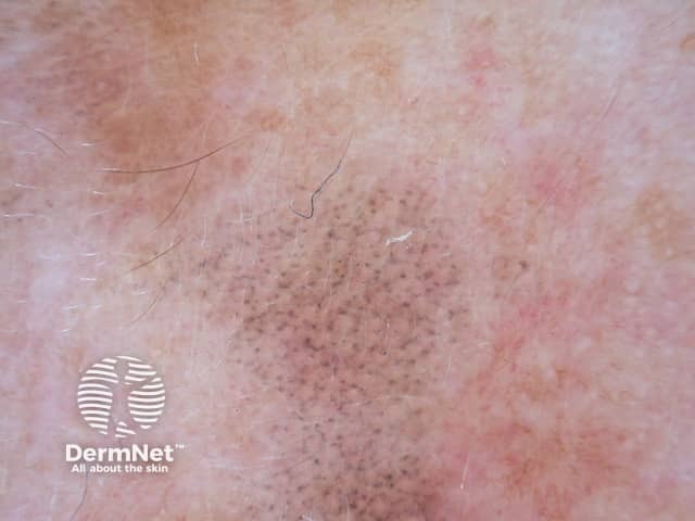
  • Dermoscopy may show remnants of pigment network, subtle blotches of brown colour, clusters of grey dots plus dotted, irregular linear and other shaped telangiectatic blood vessels
Histopathology
  • Classic variant shows epidermal acanthosis with a band-like lichenoid lymphocytic infiltrate. Presence of epidermal parakeratosis distinguishes these lesions from typical lichen planus.
  • Bullous variant shows intraepidermal or subepidermal bullous cavities with dense lymphocytic infiltrate and increased number of necrotic basilar layer keratinocytes.
  • Atypical variant shows similar histology to classic type with scattered enlarged CD-3, CD-30 (+) lymphocytes with hyperchromatic, irregular nuclei.
Early or interface subtype
Clinical features
  • Subacute lesions present for 3 months to one year
  • Erythematous to dusky-red or hyperpigmented brown lesion
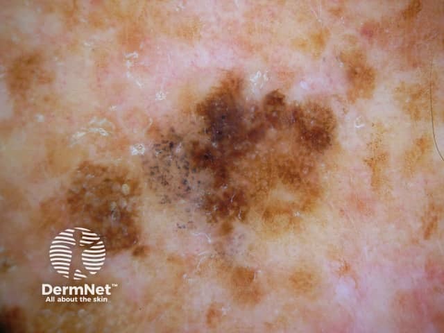
  • Depending on the age of lesion, dermoscopy may show features of a solar lentigo or flat seborrhoeic keratosis with moth-eaten borders, fingerprinting, milia-like cysts, comedo-like openings, plus small foci of melanophages (grey dots).
Histopathology
  • Single lymphocytes aligned along the dermoepidermal junction without epidermal acanthosis and adjacent lentigo
Late regressed or atrophic subtype
Clinical features
  • Lesions have been present for more than one year
  • May be violaceous papules or irregularly distributed lesions with shades of brown or grey
Histopathology
  • Epidermal atrophy with papillary dermal scarring, patchy lymphocytic infiltrates and melanin incontinence

Multiple eruptive lichenoid keratoses in sun-exposed sites are also described. Their colour varies from an initial reddish brown to a greyish purple/brown as the lesion resolves several weeks or months later.

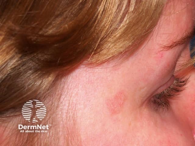
Lichenoid keratosis

How is lichenoid keratosis diagnosed?

Lichenoid keratosis may be diagnosed clinically and confirmed on dermoscopy which reveals uniform clusters of grey dots [see Annular granular pattern dermoscopy]. Depending on the stage of the lesion, there may be signs of an original pre-existing lentigo or seborrhoeic keratosis which disappear with time. Later on the grey dots also disappear, as the lesion resolves.

A skin biopsy is required if clinical examination and dermoscopy cannot differentiate between lichenoid keratosis and other solitary erythematous lesions.

Histopathology resembles lichen planus or lichenoid drug eruption, with some slight differences. Remnants of the original solar lentigo or seborrhoeic keratosis may be evident.
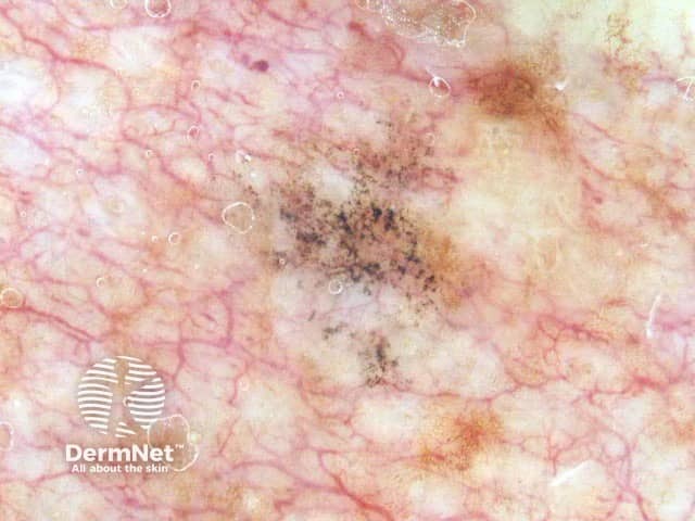
Dermoscopy of lichenoid keratosis

What is the treatment of lichenoid keratosis?

If there is any doubt about the diagnosis, dermoscopy digital images can be taken and used in follow-up a few months later.

Lichenoid keratosis can be removed if desired by liquid nitrogen, electrosurgery or curettage.

Multiple eruptive lichenoid keratoses may be effectively treated with the oral retinoid, acitretin.

What is the outcome for lichenoid keratosis?

Lichenoid keratosis is harmless and resolves spontaneously.

To date there have been no reports of lichenoid keratosis turning into malignant skin tumours.


Bibliography

  • BinJadeed H, Aljomah N, Alsubait N, Alsaif F, AlHumidi A. Lichenoid keratosis successfully treated with topical imiquimod. JAAD Case Rep. 2020;6(12):1353–5. doi:10.1016/j.jdcr.2020.10.002. PubMed Central 
  • Gori A, Oranges T, Janowska A, et al. Clinical and dermoscopic features of lichenoid keratosis: a retrospective case study. J Cutan Med Surg. 2018;22(6):561–6. doi:10.1177/1203475418786213. PubMed 
  • Maor D, Ondhia C, Yu LL, Chan JJ. Lichenoid keratosis is frequently misdiagnosed as basal cell carcinoma. Clin Exp Dermatol. 2017;42(6):663–6. doi:10.1111/ced.13178. PubMed 
  • Pitney L, Weedon D, Pitney M. Multiple lichen planus-like keratoses: lichenoid drug eruption simulant and under-recognised cause of pruritic eruptions in the elderly. Australas J Dermatol. 2016 Feb;57(1):54–6. doi: 10.1111/ajd.12288. PubMed

On DermNet

Books about skin diseases


Related information

Sign up to the newsletter