Introduction Histology Special studies Differential diagnoses
Auricle pseudocysts are rare. They are usually asymptomatic swellings which arise either spontaneously or following trauma.
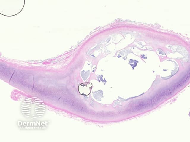
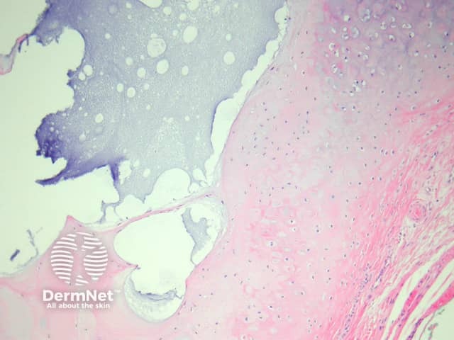
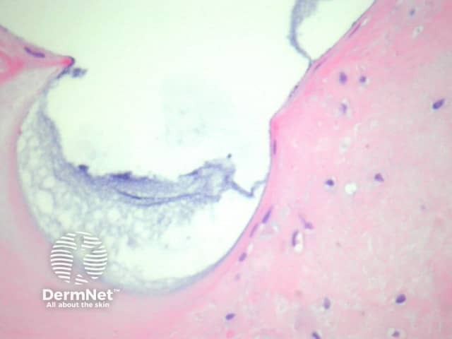
The lesion is a pseudocystic space within the substance of the cartilage (figure 1). The cartilage often shows degenerative changes and may show old haemorrhage and inflammation. There is no epithelium lining the space (hence the term “pseudocyst”). The pseudocystic space is filled with mucinous material (figures 1, 2, 3).
None are generally needed.
Epidermoid cyst – These do not arise within cartilage and are lined by a squamous epithelium.
Focal cutaneous mucinosis – This entity involves the dermis and sometimes subcutis but does not involve cartilage of the external ear.

