Introduction Which lesions should be imaged? Who should undergo photographic skin surveillance? Advantages Risks Should I have photographic skin surveillance?
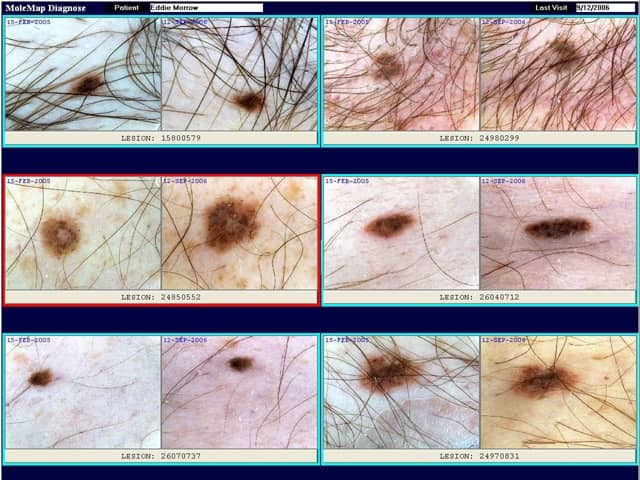
Photographic skin surveillance usually refers to a screening programme for those at high risk of malignant melanoma, for example, MoleMap New Zealand's mole mapping programme.
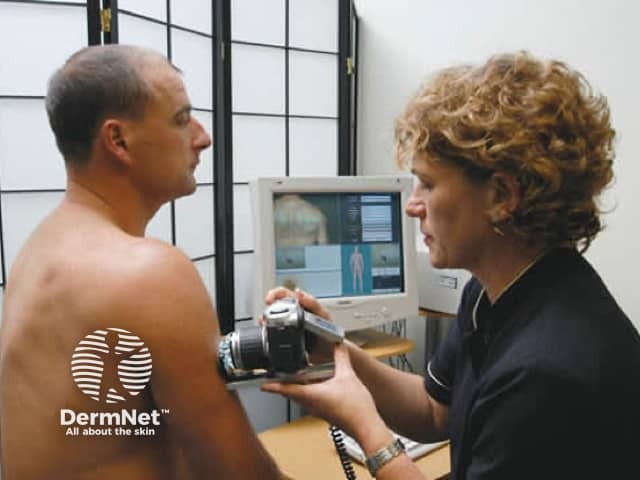
The main components are:
Digital photographic skin surveillance may include:
The patient will be asked to remove at least outer clothing. Make-up, nail varnish and jewellery should be completely removed prior to the procedure. Long hair should be tied up.
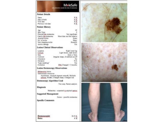
Lesions of concern are those that have features consistent with melanoma or other form of skin cancer (such as basal cell carcinoma or squamous cell carcinoma). Characteristically, skin cancers enlarge or change over periods of weeks to years.
The characteristics of melanoma are defined by the ABCDE rule and the Glasgow 7-point checklist. These are a useful guide, but may not identify early melanomas or atypical forms. Not all skin lesions with these characteristics are melanomas; many turn out to be harmless.
Letter |
Rule |
|||
|---|---|---|---|---|
A |
Asymmetry |
|||
Major features |
Minor features |
|---|---|
|
|
Nonmelanoma (or keratinocytic) skin cancers are much more common than melanoma. These usually present as growing skin lesions, that may be crusty, ulcerated or bleeding.
If you have any skin lesions that worry you because they are new, enlarging or look distinctive or unusual, ask your doctor's advice. If your doctor is also concerned, he or she may advise removal (biopsy), follow-up appointment, referral to a specialist, or photographic skin surveillance.
Photographic skin surveillance is particularly useful for individuals who have:
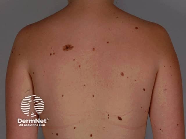
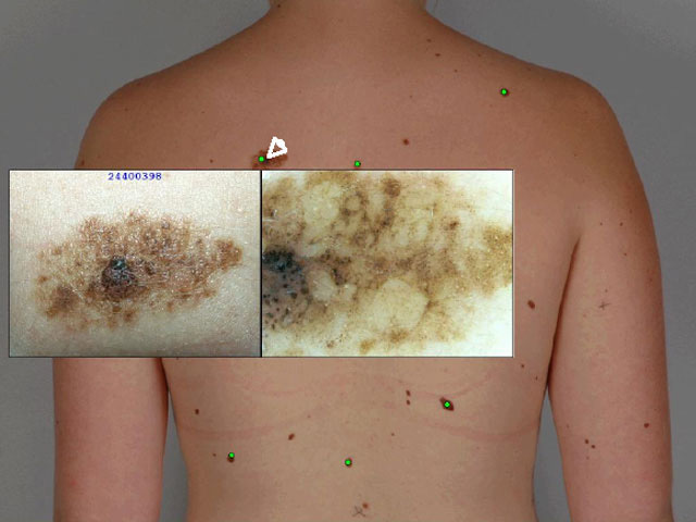
Photographic skin surveillance is most useful for pigmented moles – moles that are light to dark brown in colour (rather than pink). Accurate diagnosis depends on evaluation of the structure of the pigment.
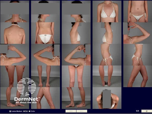
Photographic skin surveillance is intended to diagnose melanoma at the earliest possible stage, by identifying new melanocytic lesions or change in pre-existing melanocytic lesions. These features may be suspicious of melanoma if the lesion also has a disordered structure clinically or on dermatoscopy.
Compared to self skin examination or an examination by a non-specialist doctor, photographic skin surveillance has several advantages.
Clients should be aware that any screening system has risks.
If you are considering undergoing photographic skin surveillance, discuss the procedure with your own doctor. Ensure:

