DermNet provides Google Translate, a free machine translation service. Note that this may not provide an exact translation in all languages

Translate

Diabetic foot ulcer

Authors: Vanessa Ngan, Staff Writer, 2003. Revised: Dr Sashika Samaranayaka, Post Graduate Year 1, Department of Dermatology, Middlemore Hospital Auckland, New Zealand; Hon Assoc Prof Paul Jarrett, Dermatologist, Clinical Head Dermatology, Middlemore Hospital and Department of Medicine, The University of Auckland, Auckland, New Zealand. Copy edited by Gus Mitchell. January 2021.


toc-icon

What is a diabetic foot ulcer?

Diabetic foot ulcer is a skin sore with full thickness skin loss on the foot due to neuropathic and/or vascular complications in patients with type 1 or type 2 diabetes mellitus.

Who gets diabetic foot ulcer?

Diabetic foot ulcer has an annual incidence of 2–6% and affects up to 34% of diabetic patients during their lifetime. Risk factors for developing a diabetic foot ulcer include:

  • Type 2 diabetes being more common than type 1
  • A duration of diabetes of at least 10 years
  • Poor diabetic control and high haemoglobin A1c
  • Being male
  • A past history of diabetic foot ulcer.

What causes diabetic foot ulcer?

Diabetic foot ulcers are caused by neuropathic and/or vascular complications of diabetes mellitus.

Neuropathic ulcer

High blood sugar levels can damage the sensory nerves resulting in a peripheral neuropathy, with altered or complete loss of sensation and an inability to feel pain. Peripheral neuropathy develops in approximately 50% of adults with diabetes, increasing the risk of injury to the feet from pressure, cuts, or bruises.

Vascular ulcer

Blood vessels can also be damaged by long-standing high blood sugar levels, decreasing blood flow to the feet (ischaemia) and/or skin (microangiopathy). This can result in poor wound healing.

What are the clinical features of diabetic foot ulcer?

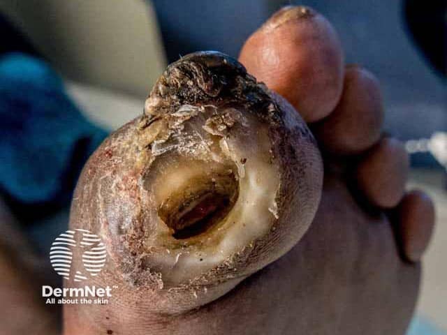
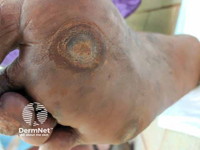
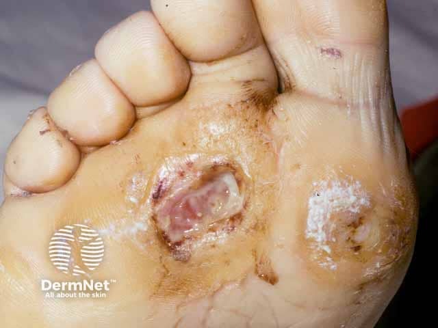
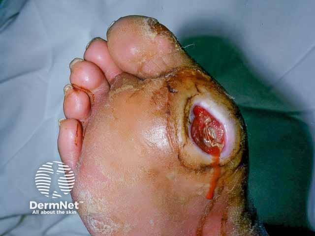
A diabetic foot ulcer is a skin sore with full thickness skin loss often preceded by a haemorrhagic subepidermal blister. The ulcer typically develops within a callosity on a pressure site, with a circular punched out appearance. It is often painless, leading to a delay in presentation to a health professional. Tissue around the ulcer may become black, and gangrene may develop. Pedal pulses may be absent and reduced sensation can be demonstrated.
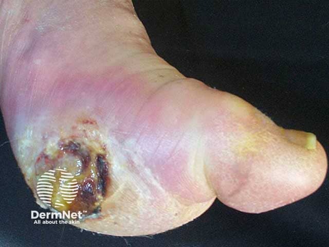
Diabetic foot ulcer

The severity of a diabetic foot ulcer can be graded and staged. There are many different classification systems. The University of Texas (UT) classification is a widely used, validated system (Table 1).

Table 1. University of Texas (UT) classification of diabetic foot ulcer

UT Grade UT Stage
0: Pre- or post-ulcerative or healed wound A: No infection or ischaemia
1: Superficial wound not involving tendon, capsule, or bone B: Infection present
2: Wound penetrating to tendon or capsule C: Ischaemia present
3: Wound penetrating to bone or joint D: Infection and ischaemia present

Grading foot ulcer

What are the complications of diabetic foot ulcer?

Diabetic foot ulcer is particularly prone to secondary infection resulting in:

  • Wound infection
  • Cellulitis
  • Osteomyelitis.

How is diabetic foot ulcer diagnosed?

Diabetic foot ulcer is a clinical diagnosis of a painless foot ulcer in a patient with a long history of poorly controlled diabetes mellitus.

Investigations may include:

  • Swabs for secondary infection
  • X-rays for bone involvement
  • Angiography.

What is the differential diagnosis for diabetic foot ulcer?

What is the treatment for diabetic foot ulcer?

Prevention of diabetic foot ulcer

  • Optimise diabetes control to reduce neuropathic and vascular complications
  • Smoking cessation
  • Regular examination of the at-risk foot, and careful toenail trimming
  • Prompt treatment of non-ulcerative conditions such as tinea pedis or cracked heels
  • Appropriate footwear — properly fitting soft shoes or made-to-measure insoles
  • Exercise and physiotherapy
  • Education of patient, family, and healthcare providers

General measures

  • Multidisciplinary care — may include endocrinologist, podiatrist, diabetes education nurse, wound care nurse, neurologist, vascular surgeon, and/or orthopaedic surgeon
  • Pressure reduction — appropriate footwear, crutches
  • Control of blood sugar levels
  • Prevention of secondary infection

Specific measures

  • Treatment of secondary infection
  • Ulcer debridement
  • Dressings
  • Adjuvant therapies — negative pressure wound therapy, ulcer excision and grafting, topical or hyperbaric oxygen, human growth factors, and/or amputation

What is the outcome for diabetic foot ulcer?

Diabetic foot ulcer may:

  • Heal over weeks to months
  • Re-ulcerate — recurrence rate is 40% within one year, 60% by three years, and 65% at five years
  • Result in lower extremity amputation in 14–24%.

The five-year mortality rate has been estimated to be 42%.


Bibliography

  • Armstrong DG, Boulton AJM, Bus SA. Diabetic foot ulcers and their recurrence. N Engl J Med. 2017;376(24):2367–75. doi:10.1056/NEJMra1615439. PubMed
  • Boulton AJM, Armstrong DG, Kirsner RS, et al. Diagnosis and management of diabetic foot complications. Arlington (VA): American Diabetes Association; October 2018. PubMed
  • Bourke J. Skin disorders in diabetes mellitus. In: Griffiths C, Barker J, Bleiker T, Chalmers R, Creamer D (eds) Rook’s Textbook of Dermatology, 9th edn. Wiley Blackwell, 2016: p64.2.
  • Everett E, Mathioudakis N. Update on management of diabetic foot ulcers. Ann N Y Acad Sci. 2018;1411(1):153–65. doi:10.1111/nyas.13569. PubMed
  • Hicks CW, Selvin E. Epidemiology of peripheral neuropathy and lower extremity disease in diabetes. Curr Diab Rep. 2019;19(10):86. doi:10.1007/s11892-019-1212-8. PubMed
  • Oyibo SO, Jude EB, Tarawneh I, Nguyen HC, Harkless LB, Boulton AJ. A comparison of two diabetic foot ulcer classification systems: the Wagner and the University of Texas wound classification systems. Diabetes Care. 2001;24(1):84–8. doi:10.2337/diacare.24.1.84. PubMed
  • Robinson TE, Kenealy T, Garrett M, Bramley D, Drury PL, Elley CR. Ethnicity and risk of lower limb amputation in people with Type 2 diabetes: a prospective cohort study. Diabet Med. 2016;33(1):55–61. doi:10.1111/dme.12807. PubMed
  • Zhang P, Lu J, Jing Y, Tang S, Zhu D, Bi Y. Global epidemiology of diabetic foot ulceration: a systematic review and meta-analysis. Ann Med. 2017;49(2):106–16. doi:10.1080/07853890.2016.1231932. PubMed

On DermNet

Other websites

Books about skin diseases


Related information

Sign up to the newsletter