What are asymmetrical pigmented follicular openings?
Asymmetrical pigmented follicular openings are curved or crescent-shaped areas of pigment partially surrounding the adnexal openings of the face.
Dermoscopic features of asymmetrical pigmented follicular openings
Asymmetrical pigmented follicular openings are seen through the dermatoscope as pigmented crescents at the periphery of a follicle. They can be the same colour as the surrounding pigment as in a solar lentigo, or heavily pigmented, often with a greyish hue as seen in lentigo maligna melanoma [1].
Lesions characterised by asymmetrical pigmented follicular openings
Asymmetrical pigmented follicular openings are characteristic of:
- Lentigo maligna
- Lentigo maligna melanoma.
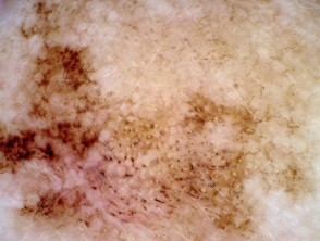
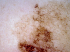
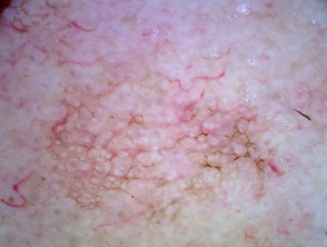
Asymmetrical pigmented follicular openings in lentigo maligna
They can also occur in:
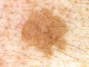
- Solar lentigo — the colour of the crescent-shaped area tends to be the same colour as the surrounding area.
- Pigmented actinic keratosis.
Although asymmetrical pigmented follicular openings are often said to be a clue to melanoma in situ, the clue actually has poor specificity [2].
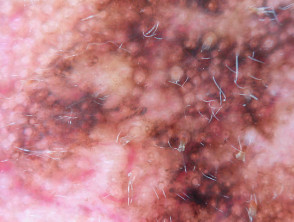
Asymmetrical pigmented follicular openings in other lesions
What is the histological explanation for asymmetrical pigmented follicular openings?
Asymmetrical pigmented follicular openings are due to abnormal melanophages spread asymmetrically up the hair follicle. In lentigo maligna, abnormal melanocytes may arise from stem cells in the follicular infundibulum.