Lipoid proteinosis is a rare autosomal recessive disease caused by mutations in the extracellular matrix protein 1 gene (ECM1).
Histology of lipoid proteinosis
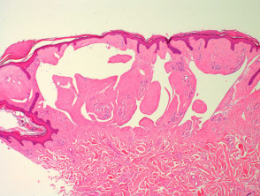
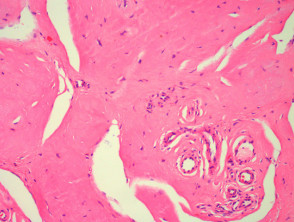
In lipoid proteinosis, sections show deposition of an eosinophilic homogenous material in the dermis (figures 1, 2). The overlying epidermis may be papillomatous (figure 1). The material surrounds blood vessels and adnexa (figure 2).
Lipoid proteinosis pathology
Special studies for lipoid proteinosis
The eosinophilic material is strongly positive with PAS and weakly positive or negative for congo red and thioflavine-T.
Differential diagnosis of lipoid proteinosis pathology
Colloid milium and amyloidosis – Both these conditions show accumulation of a similar eosinophilic material. These should both be congo red positive. Clinical history can be helpful to exclude colloid milium. Immunohistochemical studies for keratin and other amyloid-associated proteins can be helpful if amyloidosis is suspected.