Main menu
Common skin conditions

NEWS
Join DermNet PRO
Read more
Quick links
Created 2008.
Blisters are accumulations of fluid within or under the epidermis. Diagnosis depends on the site of the intercellular split as shown in the table below.
| Site of blister | Characteristics | Differential diagnosis | Image |
|---|---|---|---|
| Subcorneal | Very thin roof breaks easily | Impetigo, miliaria, SSSS |  Miliaria |
| Intra-epidermal | Thin roof ruptures to leave denuded surface | Acute eczema, varicella, herpes simplex, pemphigus |  Eczema |
| Subepidermal | Tense roof often remain intact | Bullous pemphigoid, dermatitis herpetiformis, erythema multiforme, TEN, friction blisters |  Systemic lupus erythematosus |
SSSS: Staphylococcal scalded skin syndrome
TEN: Toxic epidermal necrolysis
Epidermolysis bullosa (EB) refers to a group of inherited disorders in which there are mutations in specific keratin proteins (EB simplex), hemidesmosomes (junctional EB), anchoring filaments or type VII collagen (dystrophic EB). Minor trauma results in blisters and erosions, the split site and severity depending on the specific defect.
Epidermolysis bullosa Epidermolysis bullosa Epidermolysis bullosa 


There are at least 9 distinct immunobullous diseases due to autoantibodies directed at differing components of the desmosome complex. Skin biopsy and direct immunofluorescence are diagnostic and there may be detectable circulating skin antibodies. High dose corticosteroids and immunosuppressive medication may be required for control.
Bullous pemphigoid is the most common immunobullous disease and affects the elderly. Early signs include various subacute itchy rashes on any site, particularly the flexures (submamammary, inguinal):
Eventually (days to weeks) the plaques evolve into subepidermal bullae, or these may arise from apparently normal skin.
Purulent lesions may or may not indicate secondary infection (staphylococcal and/or streptococcal), and in such cases spreading erythema may or may not be due to cellulitis.

bullous pemphigoid

bullous pemphigoid

bullous pemphigoid

bullous pemphigoid

bullous pemphigoid

bullous pemphigoid
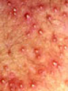
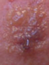
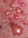
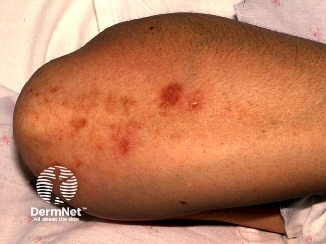
Dermatitis herpetiformis mostly affects young adults but may present at any age as a chronic prurigo. It mainly affects scalp, elbows, buttocks, knees and shoulders. Diagnosis is made by skin biopsy and rapid improvement on dapsone. Signs include:
Despite distressing itch, signs may be quite subtle (hence few photographs available!).
Dermatitis herpetiformis (DH) is associated with gluten-sensitive enteropathy in most cases (at least 85%) although this may be asymptomatic. Symptoms (in 10%) include:

Dermatitis herpetiformis

Dermatitis herpetiformis

Dermatitis herpetiformis
Pemphigus has at least 7 subtypes caused by pathogenic IgG antibodies to intraepidermal cell adhesion molecules. It is diagnosed by clinical presentation and skin biopsy. The most common subtypes are:
Pemphigus vulgaris is potentially fatal; it usually presents with acute or subacute extensive oral ulceration followed by widespread cutaneous denudation. It appears to be particularly common in middle-aged patients from the Indian subcontinent.
Pemphigus foliaceus affects the elderly and presents with erosions affecting primarily seborrhoeic areas (scalp, face, chest). It is much less severe than pemphigus vulgaris.
Pemphigus may be precipitated by:

Oral pemphigus

Pemphigus vulgaris

Pemphigus foliaceus

Paraneoplastic pemphigus

Lip erosions in paraneoplastic pemphigus (PNP-patient1)

Scalp crusting and erosions in paraneoplastic pemphigus (PNP-patient1)
Other rare immunobullous diseases include:

Pemphigoid gestationis

Cicatricial pemphigoid

Epidermolysis bullosa acquisita

Linear IgA dermatosis
Skin biopsy and direct immunofluorescence (DIF) are crucial for diagnosis of immunobullous diseases.
| Disease | Biopsy features | DIF |
|---|---|---|
| Bullous pemphigoid |
|
Linear IgG, C3 in BMZ (lamina lucida) |
| Dermatitis herpetiformis |
|
Granular IgA in tips of papillae |
| Pemphigus vulgaris |
|
IgG within suprabasal intercellular spaces |
| Pemphigus foliaceus |
|
IgG within upper epidermal intercellular spaces |
| Paraneoplastic pemphigus |
|
IgG within epidermal intercellular spaces and BMZ |
| Pemphigoid gestationis |
|
Linear C3 in BMZ (lamina lucida) |
| Cicatricial pemphigoid |
|
Linear IgG, C3 in BMZ (lamina lucida) |
| Epidermolysis bullosa acquisita |
|
Linear IgG, C3 in BMZ (upper dermis) |
| Linear IgA dermatosis |
|
Linear IgA, C3 in BMZ |
BMZ: basement membrane zone
EM: electron microscopy
Specific immunoreactive proteins detected using histochemistry on skin biopsies are beyond the scope of this article, and they are not evaluated in routine clinical practice.
There may be circulating ‘skin antibodies’ in immunobullous diseases. These are detected by indirect immunofluoresce of serum.
Additional investigations to detect gluten enteropathy should include:
Control of immunobullous diseases can be very challenging and is best left to a dermatologist. Maintenance therapy is usually required for years if not lifelong and is likely to result in significant complications.
Describe the adverse effects of dapsone.
Information for patients
See the DermNet bookstore