Introduction
Demographics
Causes
Clinical features
Types of BCC
Complications
Diagnosis
Treatment for primary basal cell carcinoma
Treatment for advanced basal cell carcinoma
Prevention
Outlook
Basal cell carcinoma (BCC) is a common, locally invasive, keratinocyte cancer (also known as nonmelanoma cancer). It is the most common form of skin cancer. BCC is also known as rodent ulcer and basalioma. Patients with BCC often develop multiple primary tumours over time.
Risk factors for BCC include:
The cause of BCC is multifactorial.
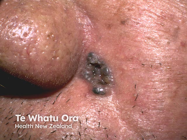
BCC is a locally invasive skin tumour. The main characteristics are:
BCC is very rarely a threat to life. A tiny proportion of BCCs grow rapidly, invade deeply, and/or metastasise to local lymph nodes.
There are several distinct clinical types of BCC, and over 20 histological growth patterns of BCC.


See more images of basal cell carcinoma.
Recurrence of BCC after initial treatment is not uncommon. Characteristics of recurrent BCC often include:


Advanced BCCs are large, often neglected tumours.
BCC is diagnosed clinically by the presence of a slowly enlarging skin lesion with typical appearance. The diagnosis and histological subtype is usually confirmed pathologically by a diagnostic biopsy or following excision.
Some typical superficial BCCs on trunk and limbs are clinically diagnosed and have non-surgical treatment without histology.
The treatment for a BCC depends on its type, size and location, the number to be treated, patient factors, and the preference or expertise of the doctor. Most BCCs are treated surgically. Long-term follow-up is recommended to check for new lesions and recurrence; the latter may be unnecessary if histology has reported wide clear margins.
Excision means the lesion is cut out and the skin stitched up.
Mohs micrographically controlled surgery involves examining carefully marked excised tissue under the microscope, layer by layer, to ensure complete excision.
Superficial skin surgery comprises shave, curettage, and electrocautery. It is a rapid technique using local anaesthesia and does not require sutures.
Cryotherapy is the treatment of a superficial skin lesion by freezing it, usually with liquid nitrogen.
Photodynamic therapy (PDT) refers to a technique in which BCC is treated with a photosensitising chemical, and exposed to light several hours later.
Imiquimod is an immune response modifier.
5-Fluorouracil cream is a topical cytotoxic agent.
A combination of 5-FU and topical calcipotriol, applied twice daily for 5-10 days, has proven more effective than monotherapy.
Radiotherapy or X-ray treatment can be used to treat primary BCCs or as adjunctive treatment if margins are incomplete.
Locally advanced primary, recurrent or metastatic BCC requires multidisciplinary consultation. Often a combination of treatments is used.
Targeted therapy refers to the hedgehog signalling pathway inhibitors, vismodegib and sonidegib. These drugs have some important risks and side effects.
The most important way to prevent BCC is to avoid sunburn. This is especially important in childhood and early life. Fair skinned individuals and those with a personal or family history of BCC should protect their skin from sun exposure daily, year-round and lifelong.
Oral nicotinamide (vitamin B3) in a dose of 500 mg twice daily may reduce the number and severity of BCCs.
Most BCCs are cured by treatment. Cure is most likely if treatment is undertaken when the lesion is small.
About 50% of people with BCC develop a second one within 3 years of the first. They are also at increased risk of other skin cancers, especially melanoma. Regular self-skin examinations and long-term annual skin checks by an experienced health professional are recommended.

