Main menu
Common skin conditions

NEWS
Join DermNet PRO
Read more
Quick links
Actinic keratosis — extra information
Lesions (cancerous) Lesions (benign)
Actinic keratosis
Last Reviewed: June, 2024
Authors: Dr Ian Coulson, Dermatologist, United Kingdom; Editor-in-Chief of DermNet (2024).
Previous contributors: Dr Amanda Oakley, Dermatologist, Hamilton, New Zealand, (1997); further updated December 2015.
Edited by the DermNet content department.
Introduction
Demographics
Causes
Clinical features
Complications
Diagnosis
Treatment
Prevention
Outlook
What is an actinic keratosis?
Actinic keratosis is a precancerous scaly spot found on sun-damaged skin, also known as solar keratosis. It may be considered an early form of cutaneous squamous cell carcinoma (a keratinocyte cancer).

A pink base with a hyperkeratotic top on the nasal bridge - a common site for actinic keratoses

An actinic keratosis on the nose

A rough scaly lesion on the back of the hand - a common site for an actinic keratosis
Who gets actinic keratoses?
Actinic keratoses affect people that have often lived in the tropics or subtropics and have predisposing factors such as:
- Other signs of photoageing skin
- Fair skin with a history of sunburn
- History of long hours spent outdoors for work or recreation
- Defective immune system.
However, even in temperate climates they may be common - in southern Ireland and northwest England up to 25% of people aged 60 years and over have at least one.
What causes actinic keratoses?
Actinic keratoses are the result of abnormal skin cell development due to DNA damage by short wavelength UVB.
They are more likely to appear if the immune function is poor, due to ageing, recent sun exposure, predisposing disease, or certain drugs.
What are the clinical features of actinic keratosis?
Actinic keratosis may be solitary but there are often multiple keratoses. The appearance varies.
- A flat or thickened papule, or plaque
- White or yellow; scaly, warty, or horny surface
- Skin coloured, red, or pigmented
- Tender or asymptomatic
They are sometimes graded according to their appearance:
Grade 1: Mild pink or grey marks with slight scale and gritty to touch
Grade 2: Moderate thicker hyperkeratosis and easily detected
Grade 3: Severe hyperkeratotic thick keratin
Grade 4: Confluent areas several centimetres with a range of the above grades of AK
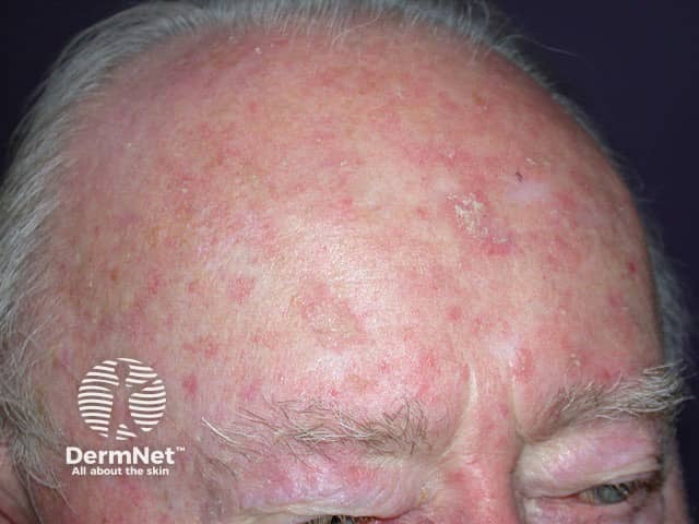
Actinic keratoses are very common on sites repeatedly exposed to the sun, especially the backs of the hands and the face, most often affecting the ears, nose, cheeks, upper lip, vermilion of the lower lip, temples, forehead, and balding scalp. In severely chronically sun-damaged individuals, they may also be found on the upper trunk, upper and lower limbs, and dorsum of feet.

Field change over the forehead with multiple superficial actinic keratoses

Thicker actinic keratoses on the dorsal hand

Hyperkeratotic actinic keratoses on the forehead
Many more images of actinic keratoses ...
- Keratoses on the nose
- Keratoses on the face
- Keratoses on the scalp
- Keratoses on the hands
- Keratoses on the legs
- Keratoses treated with imiquimod
Complications of actinic keratoses
The main concern is that actinic keratoses indicate an increased risk of developing cutaneous squamous cell carcinoma. It is rare for a solitary actinic keratosis to evolve to squamous cell carcinoma (SCC), but the risk of SCC occurring at some stage in a patient with more than 10 actinic keratoses is thought to be about 10 to 15%. A tender, thickened, ulcerated, or enlarging actinic keratosis is suspicious of evolution to SCC.
Cutaneous horn may arise from an underlying actinic keratosis or SCC.
Because they are sun damaged, people with actinic keratoses are also at risk of developing actinic cheilitis, basal cell carcinoma (BCC, which is more common than SCC), melanoma, and rare forms of skin cancer such as Merkel cell carcinoma.
How is an actinic keratosis diagnosed?
Actinic keratosis is usually easy to diagnose clinically or by dermoscopy (see actinic keratosis dermoscopy). Occasionally, a biopsy is necessary, for example, to exclude SCC, or if treatment fails.
What is the treatment for actinic keratoses?
Actinic keratoses are usually removed because they are unsightly or uncomfortable, or because of the risk that skin cancer may develop in them. The latter point is a matter of considerable debate, and the most robust studies (Cochrane review) did not find convincing evidence that AK treatment resulted in a prevention of invasive squamous cell carcinomas.
Treatment of an actinic keratosis requires removal of the defective skin cells. Epidermis regenerates from surrounding or follicular keratinocytes that have escaped sun damage. Some AKs will resolve with the frequent and appropriate use of sunscreens.
Tender, thickened, ulcerated, or enlarging actinic keratoses should be treated aggressively. Asymptomatic flat keratoses may not require active treatment but should be kept under observation.
Physical treatments
Physical treatments are used to destroy individual keratoses that are generally symptomatic or have a thick hard surface scale. The lesions may recur in time, in which case they may be retreated by the same or a different method.
Cryotherapy using liquid nitrogen
Liquid nitrogen spray is required to achieve adequate depth and duration of the freeze. This varies according to lesion location, width, and thickness. Healing varies from 5–10 days on the face, 3–4 weeks on the hands, and 6 weeks or longer on the legs. A light freeze for a superficial actinic keratosis usually leaves no mark, but longer freeze times result in hypopigmentation or scar.
Shave, curettage and electrocautery
Shave, curettage (scraping with a sharp instrument) and electrocautery (burning) may be necessary to remove a cutaneous horn or hypertrophic actinic keratosis. Healing of the wound takes several weeks or longer, depending on the body site. The specimen is sent for histological examination.
Excision
Excision ensures the actinic keratosis has been completely removed, which should be confirmed by pathology. The surgical wound is sutured (stitched). The sutures are removed after a few days, the time depending on the size and location of the lesion. The procedure leaves a permanent scar.
Field treatments
Creams are used to treat areas of sun damage and flat actinic keratoses, sometimes after physical treatments have been carried out. Field treatments are most effective on facial skin. Pretreatment with keratolytics (such as urea cream, salicylic acid ointment or topical retinoid), and thorough skin cleansing improves response rates. Results are variable and the course of treatment may need repeating from time to time. With the exception of diclofenac gel, field treatments all result in local inflammatory reactions such as redness, blistering and discomfort for a varying length of time.
- Diclofenac is more often used as an anti-inflammatory drug. A special gel formulation (3%) in a base containing sodium hyaluronate) is applied twice daily for 3 months, it is fairly well tolerated in the treatment of actinic keratoses, but less effective than the other options listed here.
- Piroxicam - similar to diclofenac.
- 5-Fluorouracil is a cytotoxic agent. The cream formulation is applied once or twice daily for 2 to 8 weeks. Its effect may be enhanced by calcipotriol ointment. 5-fluorouracil may be available as a combination product along with salicylic acid.
- Skin peels using trichloroacetic acid or Jessner solution.
- Imiquimod cream is an immune response modifier. It is applied 2 or 3 times weekly for 4 to 16 weeks.
- Photodynamic therapy (PDT) involves applying a photosensitiser (a porphyrin chemical such as methyl aminolevulinic acid) to the affected area prior to exposing it to a source of visible light.
- Tirbanibulin is a synthetic inhibitor of tubulin polymerisation and protein kinase signalling. This 5 day topical treatment received a product license in countries such as the USA, UK, and Germany in 2021.
- Oral retinoids have been used in organ transplant recipients and are shown to reduce the risk of SCC development in those with numerous AKs.
Prevention of actinic keratoses
Actinic keratoses are prevented by strict sun protection. If already present, actinic keratoses may improve with a very high sun protection factor (50+) broad-spectrum sunscreen applied at least daily to affected areas, year-round.
The number and severity of actinic keratoses can also be reduced by taking nicotinamide (vitamin B3) 500 mg twice daily.
What is the outlook for actinic keratoses?
Actinic keratoses may recur months or years after treatment. The same treatment can be repeated or another method used. Patients who have been treated for actinic keratoses are at risk of developing new keratoses. They are also at increased risk of other skin cancers, especially intraepidermal squamous cell carcinoma, invasive cutaneous squamous cell carcinoma, basal cell carcinoma and melanoma.
References
- Guidelines for the management of Actinic Keratoses (D de Berker, JM McGregor and BR Hughes) BJD, Vol. 156, No. 2, February 2007 (p222-230) — British Association of Dermatologists
- Cunningham TJ, Tabacchi M, Eliane JP, Tuchayi SM, Manivasagam S, Mirzaalian H, Turkoz A, Kopan R, Schaffer A, Saavedra AP, Wallendorf M, Cornelius LA, Demehri S. Randomized trial of calcipotriol combined with 5-fluorouracil for skin cancer precursor immunotherapy. J Clin Invest. 2017 Jan 3;127(1):106–16. doi: 10.1172/JCI89820. PubMed PMID: 27869649; PubMed Central PMCID: PMC5199703. PubMed
- de Berker D, McGregor JM, Mohd Mustapa MF, Exton LS, Hughes BR. British Association of Dermatologists' guidelines for the care of patients with actinic keratosis 2017. Br J Dermatol. 2017;176(1):20-43. doi:10.1111/bjd.15107 Journal
- Del Regno L, Catapano S, Di Stefani A, Cappilli S, Peris K. A Review of Existing Therapies for Actinic Keratosis: Current Status and Future Directions [published correction appears in Am J Clin Dermatol. 2022 Apr 22]. Am J Clin Dermatol. 2022;23(3):339-352. doi:10.1007/s40257-022-00674-3 Journal
- Dirschka T, Gupta G, Micali G, et al. Real-world approach to actinic keratosis management: practical treatment algorithm for office-based dermatology. J Dermatolog Treat. 2017;28(5):431-442. Journal
- Eisen DB, Asgari MM, Bennett DD, et al. Guidelines of care for the management of actinic keratosis. J Am Acad Dermatol. 2021;85(4):e209-e233. Journal
On DermNet
- Actinic cheilitis
- Actinic keratosis dermoscopy
- Skin cancer
- Intraepidermal squamous cell carcinoma (Bowen disease)
- Cutaneous squamous cell carcinoma
- Basal cell carcinoma
- Ultraviolet radiation
- Sun protection
- Sunscreens
Other websites
- Skin cancers and precancers — DermNet e-lectures [Youtube]
- Actinic Keratosis — Medscape Reference
- Patient Information Leaflets (PILs): Actinic keratoses — British Association of Dermatologists
- Actinic Keratosis — American Academy of Dermatology
