Subcutaneous fat necrosis of the newborn is a rare disorder that occurs in term or post-term neonates. Typical lesions include smooth, erythematous, subcutaneous nodules or plaques located on the cheeks, shoulders, back, buttocks, or thighs. Lesions usually develop within the first weeks of life and regress over the following weeks without treatment.
Histology of subcutaneous fat necrosis of the newborn
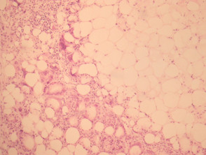
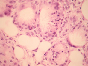
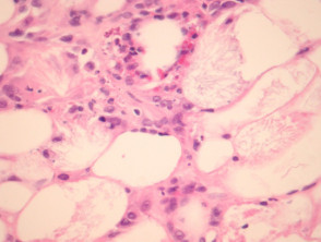
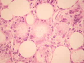
In subcutaneous fat necrosis of the newborn there is a lobular panniculitis with an infiltrate of mixed inflammatory cells. Needle-shaped clefts, in radial array, are seen in adipocytes and giant cells (figures 1–4) .
Subcutaneous fat necrosis of the newborn pathology
Special studies for sbcutaneous fat necrosis of the newborn
None are generally needed.
Differential diagnosis of subcutaneous fat necrosis of the newborn
Sclerema neonatorum: This shows identical crystals but there is minimal associated infiltrate or reaction.
Poststeroid panniculitis: This can be histologically identical and may need clinical correlation for confident distinction.