What is linear porokeratosis?
Porokeratosis is a group of skin conditions in which there is abnormal keratinisation. The skin lesions that result are reddish patches (which can be dry or atrophic) with a well-defined ridge-like border called a cornoid lamella [1]. In linear porokeratosis, the lesions are arranged in a linear formation [2].
Linear porokeratosis
Who gets linear porokeratosis?
Linear porokeratosis can be present at birth or not develop until adult life [2].
What causes linear porokeratosis?
Like other forms of porokeratosis, the cornoid lamella in linear porokeratosis is due to an expanding proliferation of unusual keratinocytes, which is thought to be due to a genetic mutation. Genetic mutations presenting in the form of mosaicism would explain the linearity and often unilateral distribution of the lesions [3].
Occasionally there is a family history of linear porokeratosis or another kind of porokeratosis, such as disseminated superficial actinic porokeratosis (DSAP), consistent with a genetic predisposition [4].
What are the clinical features of linear porokeratosis?
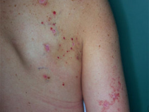
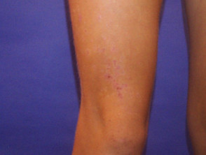
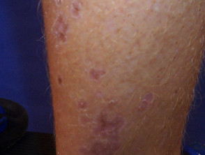
Linear porokeratosis presents as numerous grouped lesions, each with the characteristic ridge on its border and a central furrow.
They are arranged in one or more lines along a limb or on one side of the trunk, head and neck, following a dermatomal distribution (ie, along the pathway of a sensory nerve).
Localised linear porokeratosis is unilateral and is often confined to a single extremity. The generalised form of linear porokeratosis is rarer, and the lesions affect several extremities and the trunk [5].
What are the complications of linear porokeratosis?
The main complication of linear porokeratosis is skin cancer, which can develop within a linear porokeratosis lesion. This may be either a basal cell carcinoma or squamous cell carcinoma and is more likely to occur in older adults [6]. Linear porokeratosis should be monitored for malignancy.
How is linear porokeratosis diagnosed?
The diagnosis of linear porokeratosis is usually made clinically (based on the appearance), but sometimes a biopsy is needed. The biopsy should include the raised edge of the lesion. The pathology of porokeratosis is very distinct, but it may be necessary to point out the clinical features for the pathologist to find a cornoid lamella within the pathological specimen.
What is the differential diagnosis of linear porokeratosis?
Other linear lesions that should be considered when evaluating linear porokeratosis include:
- Linear epidermal naevus — this is a hyperpigmented linear birthmark. Initial flat, linear epidermal naevus becomes more warty with age. They may appear at birth or within the first few years of life.
- Lichen striatus — this is a self-limiting childhood form of blaschkoid dermatitis that presents as pink or tan raised spots that join together to form one or more dull red slightly scaly linear bands.
- Porokeratotic eccrine ostial and dermal duct naevus (PEODDN) — this is a warty birthmark that tends to occur on the hands or feet. Individual lesions are discrete papules that arrange themselves in a linear distribution.
Treatment of linear porokeratosis
There is no known cure for linear porokeratosis and treatment is generally disappointing. However, the appearance may improve with the following measures:
- 5-fluorouracil cream
- Calcipotriol cream
- Oral acitretin or isotretinoin
- Cryotherapy
- Dermabrasion
- Carbon dioxide laser ablation.
Sun protection is very important as exposure to ultraviolet radiation may result in the development of skin cancer within the linear porokeratosis.