Histology of cytomegalovirus infection
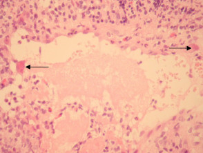
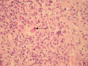
Cytomegalovirus (CMV) is a member of the family Herpesviridae. In cutaneous cytomegalovirus infection, markedly enlarged endothelial cells are seen lining small vessels (Figure 1, arrows). These are larger and eosinophilic when compared with normal or reactive endothelial cells. The hallmark histologic feature is a large intranuclear inclusion which is densely eosinophilic (Figure 2, arrow). Other cells such as fibroblasts and epithelial cells are less commonly involved.
The inclusions may be seen in the context of overlying epidermal ulceration and other non-specific inflammatory changes. Leukocytoclastic vasculitis is a described reaction pattern.
Cytomegalovirus infection pathology
Special studies for cytomegalovirus infection
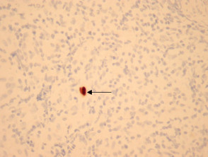
Immunohistochemical studies against CMV are a highly specific way of confirming the presence of the virus (Figure 3). PCR can also be used.
Differential diagnosis of cytomegalovirus infection pathology
Reactive changes — reactive endothelial cells can become enlarged and hyperchromatic. Dense eosinophilia (Figure 1) and the classic intranuclear inclusions (Figure 2) are not seen as a reactive phenomenon. Immunohistochemistry can be useful if needed (Figure 3).