Introduction
Neurofollicular hamartoma presents as a smooth flesh-coloured papule on the face.
Histology of neurofollicular hamartoma
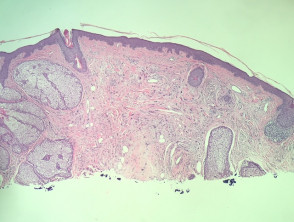
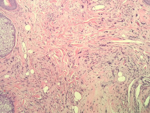
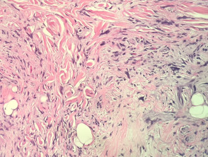
In neurofollicular hamartoma, the histopathology shows a nodular dermal spindle cell proliferation surrounded by prominent sebaceous glands. The spindle cells are plump or wavy. The sebaceous proliferation surrounds the mesenchymal component (figures 1–3).
Neurofollicular hamartoma pathology
Special studies for neurofollicular hamartoma
The spindle cells are positive for S100, CD34 and Factor XIII.
Differential diagnosis for elastofibroma
Other diagnoses to be considered include:
- Trichodiscoma — some authorities regard neurofollicular hamartoma to be a form of trichodiscoma.
- S100 is usually negative in trichodiscoma
- Neurofibroma and neurotised naevus do not usually have malformed follicular and sebaceous structures.