What is peristomal intestinal metaplasia?
Peristomal intestinal metaplasia (PIM) is a rare but serious complication affecting the skin around a gastrointestinal stoma site.
It is characterised by the histological appearance of scattered intestinal mucosa and goblet cells within the variably intact epidermis around the stoma site.
Who gets peristomal intestinal metaplasia?
PIM is rarely reported and may occur in those with an ileostomy or a colostomy.
Risk factors include:
- Any breaks or changes in the peristomal skin, which can prevent adequate adhesion of the stomal appliance
- Chronic inflammation and irritation of the peristomal skin
- Other peristomal skin conditions, most commonly peristomal dermatitis
- Effluent leakage
- Ill-fitting stomal appliances
- Peristomal hernia.
What causes peristomal intestinal metaplasia?
The exact pathogenesis is not yet understood.
Suggested causative factors include:
- Chronic inflammation of the peristomal skin
- Exposure of the peristomal skin to intestinal bile salts in the stomal effluent, which increases intestinal epithelial cell migration
- Seeding of intestinal mucosal cells into the peristomal skin during surgical procedures.
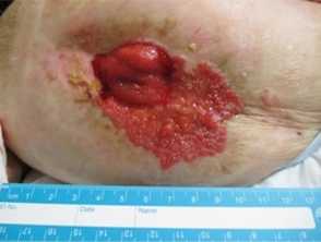
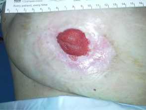
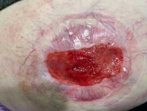
What are the clinical features of peristomal intestinal metaplasia?
PIM may present with the following clinical features:
- Painful and irritated peristomal skin
- Erosive plaque/s around the stoma site
- Peristomal ulceration
- Peristomal maceration.
How do clinical features vary in differing types of skin?
Limited case reports in the literature make it difficult to compare presentations between different ethnicities. However, to date, there have been similar presentations across different Fitzpatrick skin types.
What are the complications of peristomal intestinal metaplasia?
- Adenomas (polyps) in the peristomal skin
- Malignant transformation to adenocarcinoma
How is peristomal intestinal metaplasia diagnosed?
The diagnosis of PIM is confirmed by skin biopsy, typically shave biopsy, of the epithelium at the peripheral border of the plaque/lesion. The characteristic histological appearance of PIM is that of intestinal mucosa infiltrating the epidermis around the stoma site.
What is the differential diagnosis for peristomal intestinal metaplasia?
- Peristomal dermatitis
- Peristomal pyoderma gangrenosum
- Malignant lesion, eg, basal cell carcinoma, squamous cell carcinoma
- Peristomal overgranulation (nodular and polypoid histological changes)
- Mechanical trauma
- Peristomal cellulitis
What is the treatment for peristomal intestinal metaplasia?
General measures
- Well-fitting stoma appliances — a specialist stoma nurse will advise on the most appropriate appliance and ensure the product adheres to the peristomal skin comfortably to minimise irritation and leakage.
- Proper stoma care and education.
Specific measures
- Silver nitrate cauterisation, cryotherapy, and carbon dioxide laser treatment have been used in case reports.
- Consider sequential electrocautery under local anaesthetic monthly for 4 months, and then as indicated for any sites of recurrence.
- Revision of the stoma is sometimes found to be indicated — this is usually a collaborative decision involving the patient, surgeon, and dermatologist.
- Serial monitoring for progression to adenocarcinoma.
- Surgical excision of the carcinoma
- Chemotherapy
- Radiotherapy
- Intralesional 5-fluorouracil (5FU)
How do you prevent peristomal intestinal metaplasia?
- Proper stoma care and education by a trained professional, preferably a specialist stoma nurse.
- Well-fitting stoma appliances to minimise effluent leakage and mechanical stripping of the peristomal skin.
- Early recognition of the onset of the condition to allow for early intervention.
What is the outcome for peristomal intestinal metaplasia?
Electrocoagulation therapy has produced promising results in most case reports to date. Unfortunately, the rate of transformation to adenocarcinoma is not known.