What is a respiratory disease?
A respiratory disease refers to any condition that affects the upper respiratory tract, trachea, lungs, and the nerves and muscles associated with breathing. The correct recognition of cutaneous signs of respiratory disease can aid clinicians in diagnosing and treating these potentially life-threatening diseases.
What are the skin signs of respiratory disease?
Cyanosis
Cyanosis is a blue discolouration of the skin and mucous membranes. It is seen in patients with more than 5 g/dL of desaturated haemoglobin. Cyanosis may be:
- Central — on the lips and tongue; relates to a circulatory or respiratory problem associated with poor blood oxygenation in the lungs
- Peripheral — on the extremities or fingers; due to oxygen-depleted peripheral blood.
Cyanosis


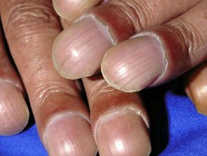
Nail clubbing
Nail clubbing is a deformity of the nails characterised by:
- Loss of the Lovibond angle (the normal < 165° angle between the nail bed and the nail fold)
- Thickening of the end of the finger
- An increase in nail fold convexity
- A soft boggy nail texture [1].
Clubbing is associated with a wide range of diseases, including the following respiratory diseases:
- Lung cancer
- Interstitial lung disease; most commonly, fibrosing alveolitis (inflammation of the alveoli in the lungs)
- Tuberculosis
- Cystic fibrosis
- Suppurative (pus-forming) lung disease, such as lung abscess, empyema (pus buildup in the pleural cavity), and bronchiectasis (damage to the lungs due to infections)
- Mesothelioma (a tumour of the lining of the lung)
- Arteriovenous fistula or malformation (abnormal formation or connection between arteries and veins).
Hypertrophic osteoarthropathy
Hypertrophic osteoarthropathy comprises:
- Clubbing of the fingers
- Periostitis (inflammation of the layer of connective tissue around a bone)
- Arthritis (inflammation of a joint).
Symptoms of hypertrophic osteoarthropathy may precede the diagnosis of lung cancer and are most commonly associated with non-small-cell lung cancer, particularly squamous cell carcinoma and adenocarcinoma [2]. Hypertrophic osteoarthropathy can also be secondary to a lung abscess, mesothelioma, and other disorders [1].
Clubbing


Lung cancer
Cutaneous metastases in lung cancer are found in 2–8% of cases of lung cancer [1]. The most common sites are the chest and abdominal wall, neck, and scalp. They are usually rapidly growing, hard, painless and mobile nodules. A punch biopsy of a nodule may be nonspecific or may establish the diagnosis of lung cancer.
Monitoring changes in the size of skin nodules can help assess the patient's response to chemotherapy [1]. Other cutaneous manifestations of lung cancer include:
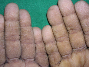
- Acanthosis nigricans
- Erythema gyratum repens
- Necrolytic migratory erythema
- Tripe palms
- Dermatomyositis
- Migratory superficial thrombophlebitis (Trousseau syndrome)
- Acquired ichthyosis
- Acquired hypertrichosis lanuginosa [3].
Cutaneous signs of lung cancer


Superior vena cava syndrome
Superior vena cava syndrome is due to obstruction of the superior vena cava, the vein that takes deoxygenated blood from the upper body to the heart. The majority of cases are due to malignancy; most commonly, non-small-cell lung cancer, small-cell lung cancer, lymphoma, and metastatic tumours [4]. Superior vena cava syndrome presents with markedly dilated veins or venules in the upper chest [5]. These occur as a result of increased collateral flow through the superficial vasculature in the chest wall.
Sarcoidosis
Sarcoidosis can be divided into two separate conditions:
- A multisystem disease, with 30% of patients having cutaneous involvement [6]
- Cutaneous sarcoidosis, in which there is no systemic involvement [7].
Cutaneous involvement is classified as either specific, in which there are non-caseating granulomas (mixed chronic inflammatory cells) on histopathology, or nonspecific, in which granulomas are absent.
Specific lesions of sarcoidosis include:
- Lupus pernio
- Macular sarcoidosis
- Plaque sarcoidosis
- Subcutaneous nodular sarcoidosis (Darier–Roussy sarcoidosis)
- Scar and tattoo infiltration.
Non–specific lesions of sarcoidosis include:
- Erythema nodosum (particularly in the early stages of sarcoidosis)
- Nummular eczema
- Erythema multiforme
- Calcinosis cutis
- Pruritus [8].
Cutaneous sarcoidosis


Cystic fibrosis
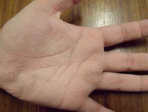
Cutaneous manifestations of cystic fibrosis may be nonspecific, but include:
- Aquagenic skin wrinkling, due to increased concentration of electrolytes in the sweat
- Cutaneous vasculitis, due to circulating antigen-antibody complexes (the antigens are bacteria, antibiotics and pancreatic enzyme supplements)
- Cystic fibrosis nutrient-deficiency dermatitis [9].
Cutaneous signs of cystic fibrosis

Granulomatosis with polyangiitis
Cutaneous manifestations of granulomatosis with polyangiitis (also known as Wegener granulomatosis) are found in 10% of patients at diagnosis and develop in 50% of patients throughout the disease [1,8].
Skin findings include:
- Necrotic ulcers, usually on the lower extremities
- Palpable purpura (raised dark spots due to small-vessel vasculitis), usually on the lower extremities
- Nodules, papules, and vesicles
- Pyoderma gangrenosum (rare)
- Raynaud phenomenon (rare)
- Mouth ulcers and gingivitis [1].
Cutaneous signs of granulomatosis with polyangiitis


Eosinophilic granulomatosis with polyangiitis
Eosinophilic granulomatosis with polyangiitis (also known as Churg-Strauss syndrome) is a rare multisystem disorder that primarily affects the lungs, skin, and peripheral nervous system. The main features are asthma, eosinophil infiltration in the lungs and blood, and small-vessel vasculitis with granulomas on biopsy.
A variety of skin lesions are seen in eosinophilic granulomatosis with polyangiitis:
- Palpable purpura, most often seen on the lower extremities
- Subcutaneous nodules, often on limbs or the scalp
- Urticaria and urticaria-like rashes
- Livedo reticularis [1].
Papules and nodules may become necrotic, starting with a central black dimple.
Vasculitis in eosinophilic granulomatosis


Pulmonary arteriovenous malformations
Pulmonary arteriovenous malformations are abnormal communications between the arteries and veins that supply the lungs. The majority of these malformations are congenital. Approximately 70% of patients with pulmonary arteriovenous malformations have associated hereditary haemorrhagic telangiectasia (also known as Osler–Weber–Rendu syndrome) in which telangiectasia is near the surface of the skin. The face, hands, feet, chest, lips, tongue, oral mucosa, and nasal mucosa are most commonly affected [1].
Alpha-1-antitrypsin deficiency
Alpha-1-antitrypsin deficiency is a genetic disorder where there is defective production of the alpha-1-antitrypsin enzyme. It primarily affects the lungs (with emphysema), liver, and skin. Cutaneous findings include recurrent ulcerative, necrotising panniculitis (inflamed subcutaneous fat with crops of painful, red subcutaneous nodules), which most noticeably occurs on the trunk and proximal extremities. These nodules typically ulcerate, releasing a clear to yellow oily fluid [1].
Fat embolism syndrome
Fat embolism syndrome typically follows fractures and trauma to the long bones or pelvis. Fat particles released into the circulation may cause:
- Respiratory distress
- Altered mental status
- Thrombocytopenia.
The classic cutaneous manifestation of fat embolism syndrome is a petechial eruption with 2–3 mm purpuric macules on non-dependent portions of the body (upper chest, neck, axillae, and conjunctivae). This finding often disappears within 5–7 days, but may be key for diagnosis [10,11].
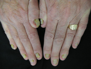
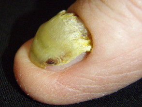
Yellow nail syndrome
Yellow nail syndrome is the triad of:
- Yellow nails
- Lymphoedema (swelling caused by blockage of the lymphatic system)
- Pleural effusions (fluid in the space surrounding the lungs) [8].
Nail changes that are usually the first features of yellow nail syndrome include:
- Onycholysis (separation of the nail from the nail bed)
- Transverse ridging on a smooth base
- Loss of the cuticle
- Slow growth (less than half the rate of healthy nails) [7].
The nails are occasionally green in colour. Lymphoedema associated with yellow nail syndrome typically affects the legs and is symmetrical and non-pitting.
Yellow nail syndrome

Birt–Hogg–Dubé syndrome
Birt–Hogg–Dubé syndrome is an autosomal-dominant inherited disease characterised by skin tumours, pneumothorax (lung collapse), kidney tumours, and lung cysts [12].
Cutaneous lesions include:
- Fibrofolliculomas (tumour developing in the hair follicles)
- Trichodiscomas (tumour of the hair disc)
- Acrochordons (skin tags).
Fibrofolliculomas and trichodiscomas are most frequently found on the head, face, and upper body.
- They are asymptomatic, pale yellow, slightly raised, dome-shaped papules measuring 2–4 mm in diameter.
- There may be few or many hundreds of lesions.
- They usually develop at around 30–40 years of age [1].
Trichofolliculomas in Birt-Hogg-Dubé syndrome


