DermNet provides Google Translate, a free machine translation service. Note that this may not provide an exact translation in all languages
Home Dermoscopy course images
Dermoscopy course images
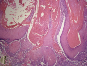
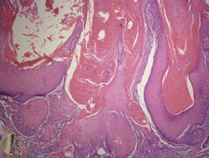
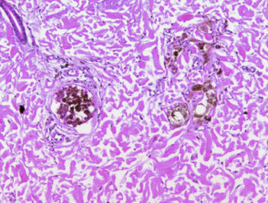
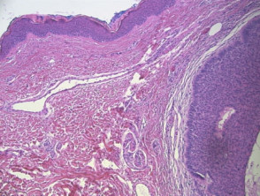
Histology of seborrhoeic keratosis
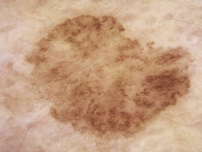
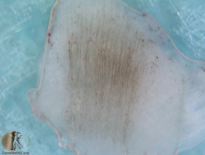
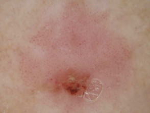
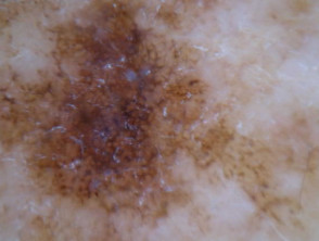
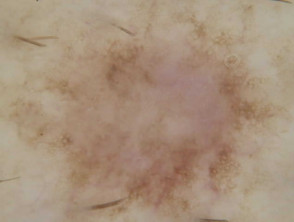
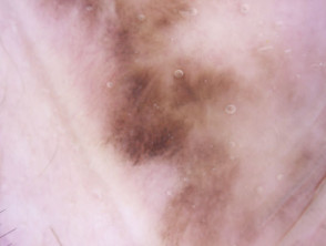
Dermatoscopy of solar lentigo
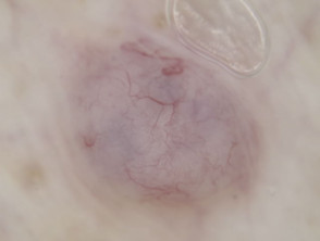
Dermatoscopy of papillomatous dermal melanocytic naevus
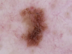
Dermoscopy of acral melanoma

white structures69c
Dermatoscopy of nail matrix melanoma
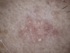
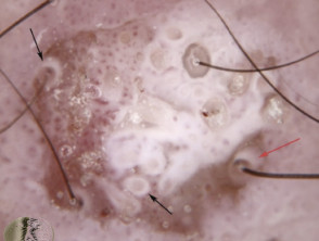
White circles on dermatoscopy of SCC
Dermatoscopy of sebaceous hyperplasia
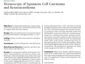
Abstract: Rosendahl C et al Dermatoscopy of squamous cell carcinoma and keratoacanthoma. Arch Dermatol 2012; 148: 1386-92
Histology of dermatofibroma
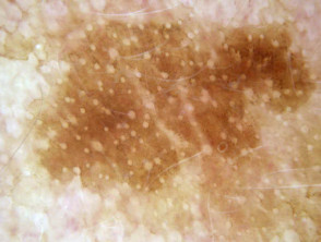
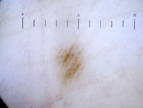
Dermatoscopy of solar lentigo
Histology of seborrhoeic keratosis
Histopathology of basal cell carcinoma
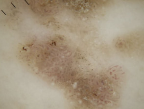
Ridge pattern, parallel
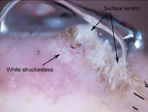
White structures and surface keratin in squamous cell carcinoma dermoscopy
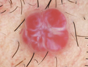
Red clods separated by white lines in pyogenic granuloma
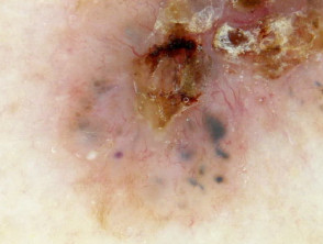
Dermatoscopy of pigmented intraepithelial carcinoma
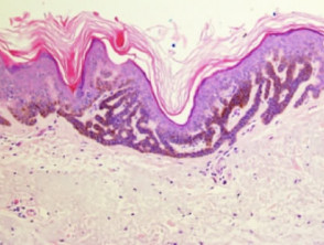
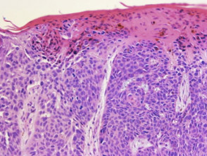
Histopathology of intraepithelial carcinoma
In-vivo dermatoscopy of pigmented nail plate
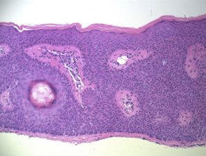
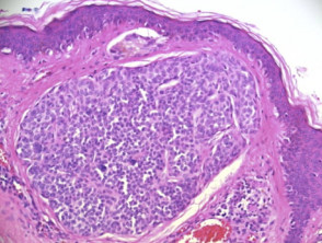
Histopathology of melanocytic naevus
Histopathology of dysplastic naevus
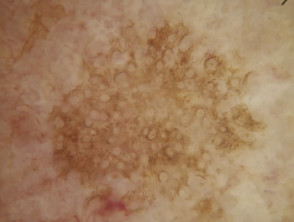
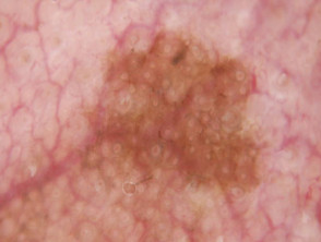
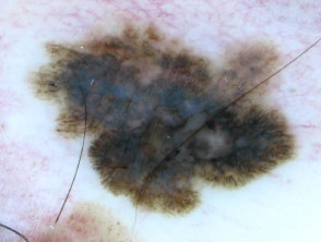
Seborrhoeic keratosis dermoscopy
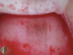
Dermatoscopic image of labial melanotic macule
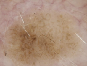
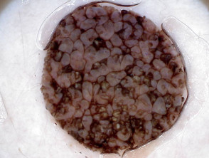
Dermatoscopy of seborrhoeic keratosis
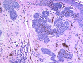
Histopathology of SCC
Structureless blue colour of blue naevus on dermatoscopy
Histopathology of basal cell carcinoma
white structures69d
Histopathology of melanocytic naevus
Histopathology of SCC
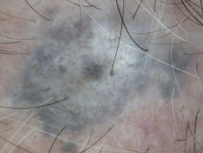
Dermatoscopy of basal cell carcinoma
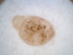
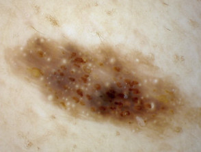
Dermatoscopic image of ink spot lentigo
White structureless areas and white circles in squamous cell carcinoma dermoscopy
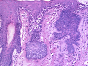
Histology of melanoma
Dermatoscopy of intraepithelial carcinoma
Histology of melanoma
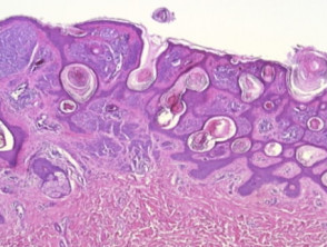
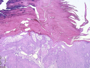
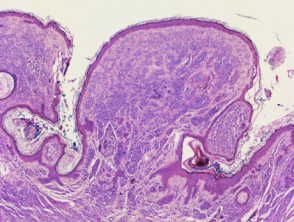
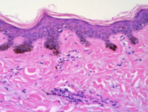
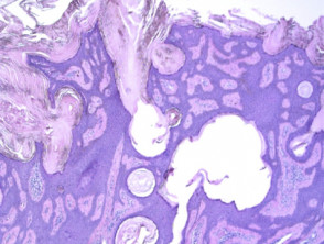
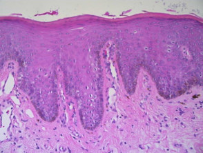
Histology of seborrhoeic keratosis
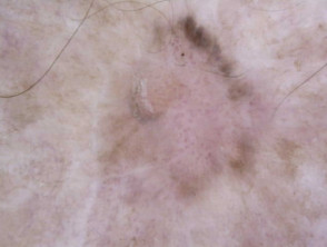
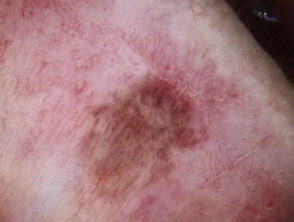
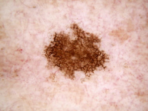
Dermatoscopy of dysplastic naevus
Histopathology of facial melanoma in situ (lentigo maligna)
acral naevus26

White circles on dermatoscopy of SCC
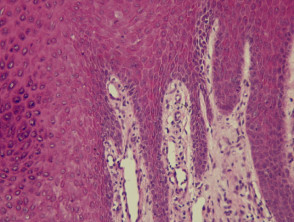
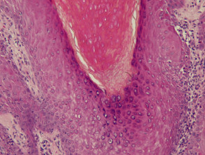
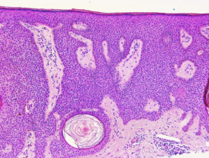
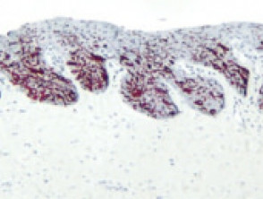
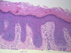
Histopathology of skin. Normal interdigitation of epidermal rete and dermal papillae.
Dermatoscopy of seborrhoeic keratosis
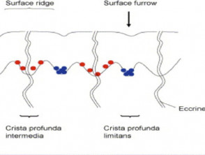
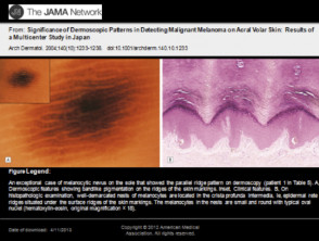
Diagram of acral volar skin
Dermatoscopy of seborrhoeic keratosis
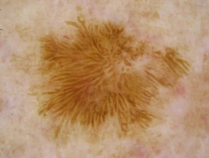
Lichen planus-like keratosis: dermatoscopy
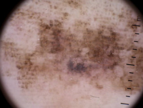
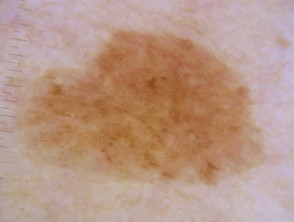
Grey circles seen in dermoscopy of a facial solar lentigo.
white structures70
Structureless blue colour of blue naevus on dermatoscopy
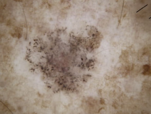
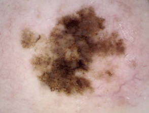
Grey circles and the 'isobar' sign in dermoscopy of a lentigo maligna
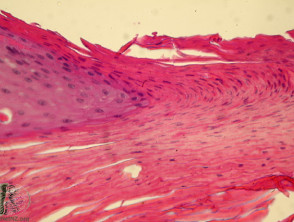
Histopathology of facial skin
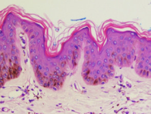
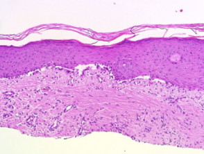
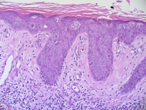
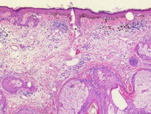
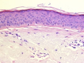
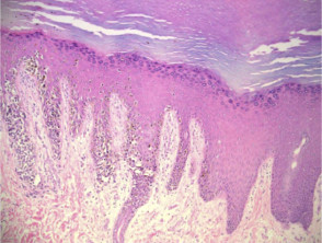
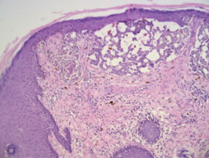
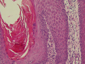
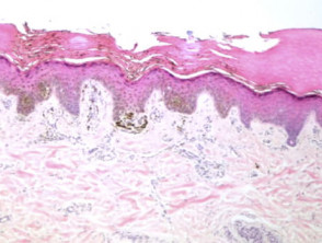
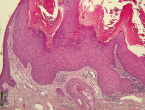
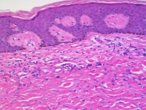
Histopathology of solar lentigo

white structures68

white structures69b
Histopathology of SCC
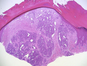
Histopathology of seborrhoeic keratosis
In-vivov dermatoscopy of pigmentation in nail bed
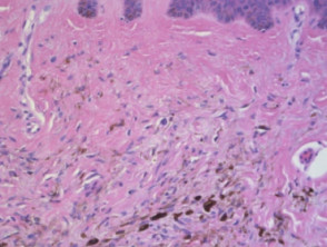
Histopathology of solar lentigo
Dermatoscopy of seborrhoeic keratosis
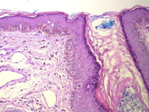
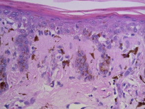
Histology of acral melanoma
Histopathology of melanocytic naevus
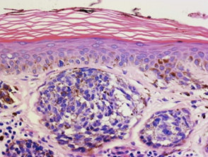
Histopathology of nests of pigmented melanocytes
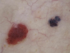
Red and blue clods on dermatoscopy of haemangioma
Lichen planus-like keratosis: dermatoscopy

white structures67b
Histopathology of seborrhoeic keratosis
White circles on dermatoscopy of SCC
Histopathology of melanocytic naevus
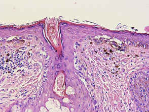
Histology of nail matrix melanoma
Histopathology of basal cell carcinoma
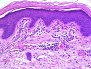
Histology of acral melanocytic naevus
Histology of melanoma
Dermatoscopy of dermal melanocytic naevus
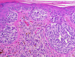
Histology of melanoma

white structures67c
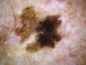
Dermatoscopy of melanoma with thick lines
Histopathology of skin showing papillary vessels
Histopathology of SCC
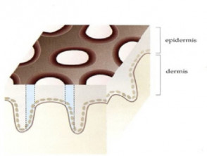
diagram pores4b
Dermoscopy of basal cell carcinoma showing blue clods
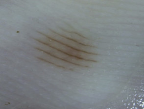
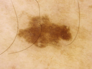
Parallel furrow pattern
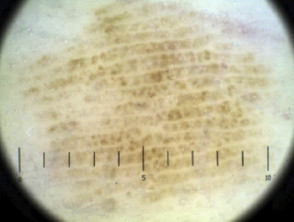
Ridge pattern, parallel
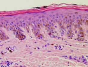
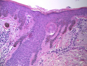
Histopathology of pigmented actinic keratosis
Histology of dermatofibroma
Dermatoscopy of dermatofibroma
Histopathology of haemangioma
Dermatoscopy of melanoma with thick lines
Dermatoscopy of solar lentigo
Dermatoscopic image of genital melanotic macule
Blue naevus
The histologic basis for reticular / branched lines in dermatoscopy
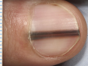
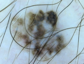
Melanonychia due to melanoma
Circles on dermatoscopy of facial melanoma in situ, lentigo maligna
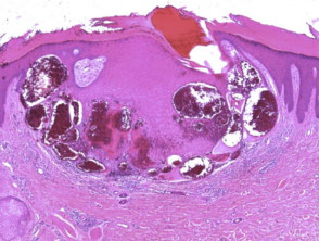
Histopathology of pyogenic granuloma
Dermatoscopic image of ink spot lentigo