What is atypical fibroxanthoma?
Atypical fibroxanthoma (AFX) is a dermal spindle-cell tumour that typically occurs on the head and neck of sun damaged older people. The tumour-like growth should be considered a type of skin cancer but it may behave in a benign fashion.
A rare type of atypical fibroxanthoma occurs in younger patients on parts of the body that are not normally overexposed to the sun. These tumours are usually found on the trunk and extremities and tend to be larger and slower growing.
Atypical fibroxanthoma
What causes atypical fibroxanthoma?
The development of atypical fibroxanthomas is associated with ageing, sun exposure (ultraviolet radiation) and/or ionising radiation (X-rays). Both forms of radiation can cause abnormal growth of atypical spindle cells. These are believed to come from fibrous cells in the dermis or from epidermal keratinocytes.
Who gets atypical fibroxanthoma?
Reports have shown an increased incidence of atypical fibroxanthoma in patients with acquired immune deficiency syndrome (AIDS) and in patients who are immune suppressed, for example because of organ transplantation.
Atypical fibroxanthoma affects both sexes equally, with a mean age of 69 years at diagnosis.
What are the clinical features of atypical fibroxanthoma?
Atypical fibroxanthoma often appears in areas that have received excessive sun exposure, usually around the scalp, ears, nose, cheeks, and back of the neck, or in areas where individuals may have previously received radiotherapy treatment. They have also been reported to occur on the trunk, extremities, and in sun-protected areas.
- A solitary tumour or multiple tumours may occur.
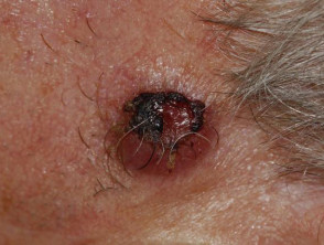
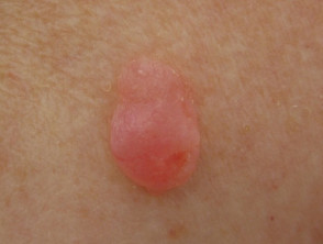
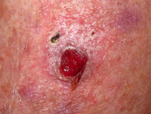
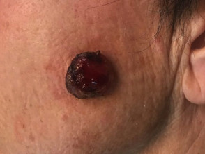
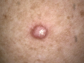
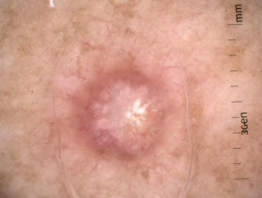
- Typically, AFX is a red, juicy, dome-shaped nodule that may be bleeding, ulcerated or crusted.
- The tumour starts off as a small nodule that grows quickly over 6 months to a size of about 2–3 cm.
- Four times as many tumours are diagnosed on the head and neck as are diagnosed in other body sites.
Although rare, cutaneous metastases from atypical fibroxanthoma have been reported.
Atypical fibroxanthoma
How is atypical fibroxanthoma diagnosed?
Because atypical fibroxanthoma can look like other skin cancers, it is usually diagnosed by a pathologist after a skin biopsy or excision.
The diagnosis depends on finding large, pleomorphic, fibrocytic, spindle-shaped, anaplastic tumour cells haphazardly arranged in the dermis. Immunohistochemistry stains should be undertaken but may be nonspecific. Cytokeratin and melanoma stains are negative.
Dermoscopic features may resemble those of basal cell carcinoma or squamous cell carcinoma.
Atypical fibroxanthoma macro + dermoscopy
What is the differential diagnosis for atypical fibroxanthoma?
Other lesions to be considered in the differential include:
- Pyogenic granuloma
- Spindle cell squamous cell carcinoma
- Spindle cell melanoma
- Superficial portion of a malignant fibrous histiocytoma
- Leiomyosarcoma.
What is the treatment of atypical fibroxanthoma?
Atypical fibroxanthoma is treated by complete surgical excision. Small lesions may be removed by curettage. Mohs micrographic surgery is becoming the treatment of choice for large or recurrent lesions, as it reliably removes the complete tumour while sparing surrounding normal healthy tissue.
What is the outcome for atypical fibroxanthoma?
Atypical fibroxanthoma rarely recurs after complete excision with clear margins. Recurrence and metastasis are more likely in people that are immunosuppressed.
How is atypical fibroxanthoma prevented?
Most cases of atypical fibroxanthoma could be prevented by avoiding excessive sun exposure. Patients are advised to follow sun protection methods when outdoors.