What is a seborrhoeic keratosis?
Seborrhoeic keratosis is a benign acanthoma comprised of epidermal keratinocytes. Clinically it presents as a sharply demarcated warty plaque with a ‘stuck-on’ appearance and greasy texture.
What is the histology of seborrhoeic keratosis?
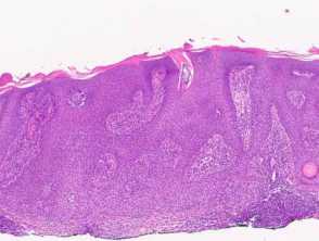
Seborrhoeic keratosis is a well-demarcated exophytic, flat or, less commonly, endophytic lesion composed of a proliferation of epidermal keratinocytes. Seborrhoeic keratosis can be recognised by a papillomatous architecture, acanthosis, hyperkeratosis, and horn cysts. The hyperkeratosis produces a characteristic loose lamellar stratum corneum. Squamous eddies may be present if particularly acanthotic.
Figure 1
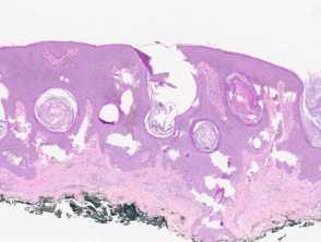
There are multiple distinct histological patterns:
- Regular acanthotic (Figure 1)
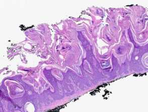
- Keratotic papillomatous (Figure 2)
- Exaggerated hyperkeratosis and papillomatosis
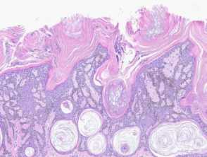
- Reticulated adenoid (Figure 3)
- Thinned rete ridges which articulate
- Small horn cysts
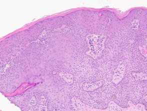
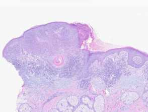
- Clonal (Figure 4)
- Nests of clear keratinocytes in the epidermis
- Irritated (Figure 5)
- Scale crust, focal squamous eddies
- Underlying lichenoid inflammation
- Pigmented (Figure 3)
- Increased epidermal pigment
- Dermal melanophages
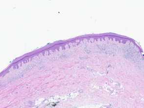
- Macular (Figure 6)
- No horn cysts
- May be difficult to distinguish from solar lentigo
- Overlap with additional features
- Pigmented, irritated, or inflamed (Figure 7)
Histological patterns of seborrhoeic keratosis
What is the differential diagnosis of seborrhoeic keratosis pathology?
- Verruca vulgaris: The keratotic papillomatous variant of seborrheic keratosis is histologically very similar to verruca vulgaris. However, verruca vulgaris shows koilocytes and inward bending rete. Horn cysts are not usually seen in verruca vulgaris [see Verruca vulgaris pathology].
- Epidermal naevus: While the histological features may be similar, the history will be of a longstanding lesion typically appearing at birth or in early childhood [see Epidermal naevus pathology].
- Confluent and reticulated papillomatosis: This presents as a netlike hyperpigmented eruption of papules and plaques so it is clinically quite distinct from a seborrheic keratosis, although the histology can be similar.