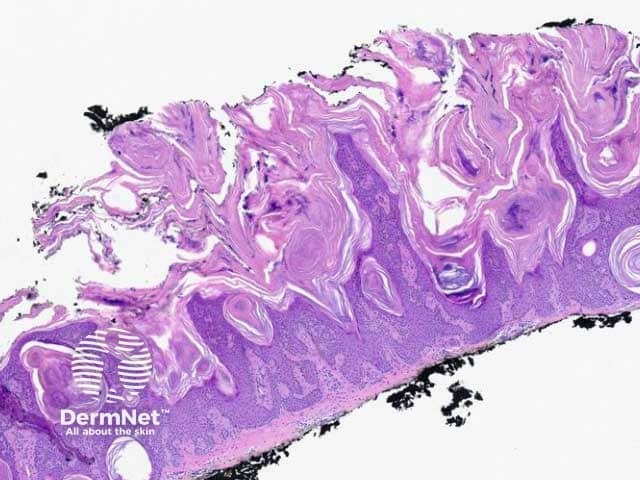
Seborrhoeic keratosis is a benign acanthoma comprised of epidermal keratinocytes. Clinically it presents as a sharply demarcated warty plaque with a ‘stuck-on’ appearance and greasy texture.
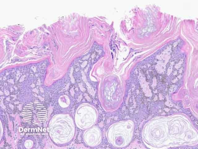
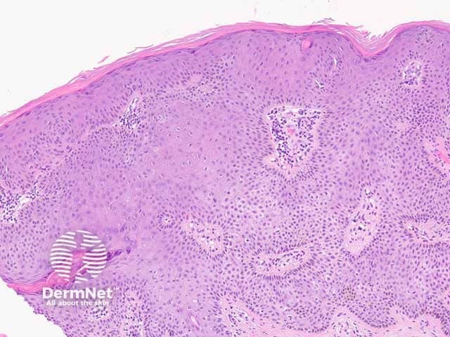
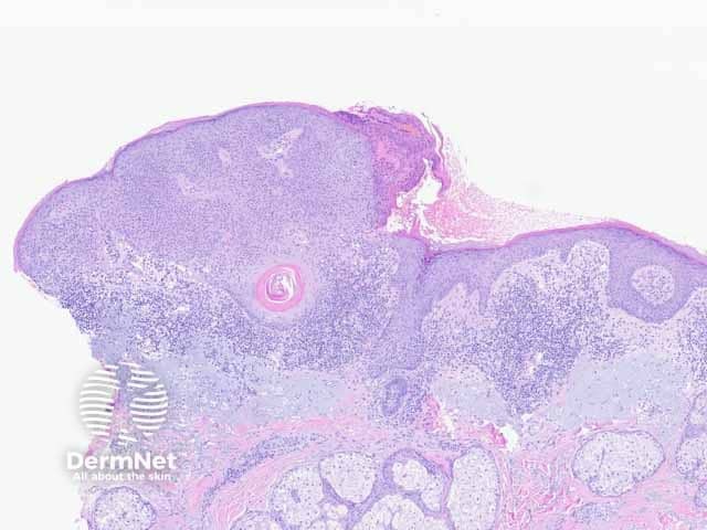
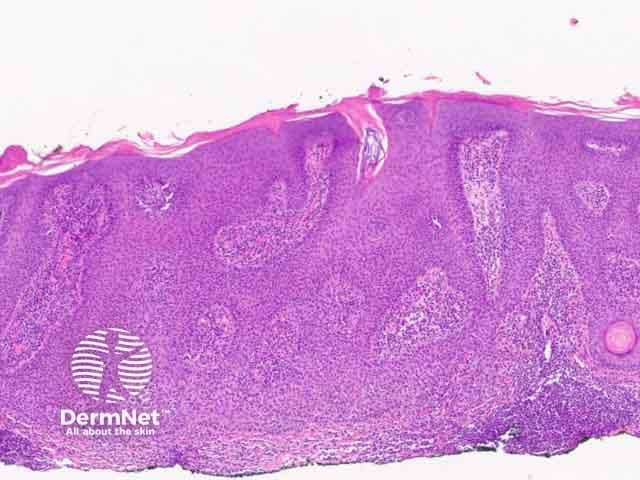
Seborrhoeic keratosis is a well-demarcated exophytic, flat or, less commonly, endophytic lesion composed of a proliferation of epidermal keratinocytes. Seborrhoeic keratosis can be recognised by a papillomatous architecture, acanthosis, hyperkeratosis, and horn cysts. The hyperkeratosis produces a characteristic loose lamellar stratum corneum. Squamous eddies may be present if particularly acanthotic.
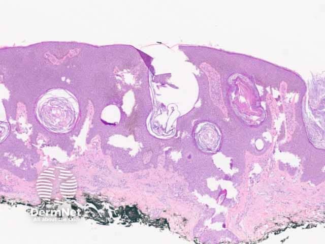
There are multiple distinct histological patterns: