Introduction
Demographics
Causes
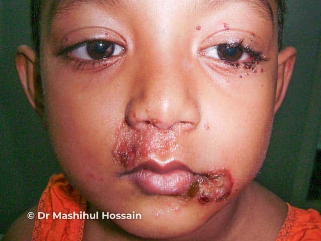
Clinical features
Variation in skin types
Complications
Diagnosis
Differential diagnoses
Treatment and prevention
Outcome
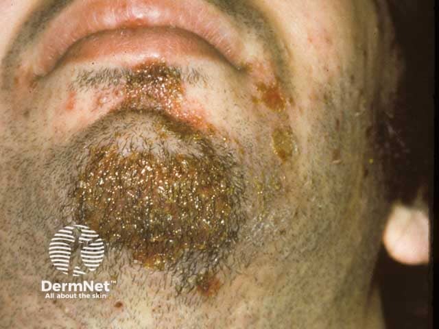
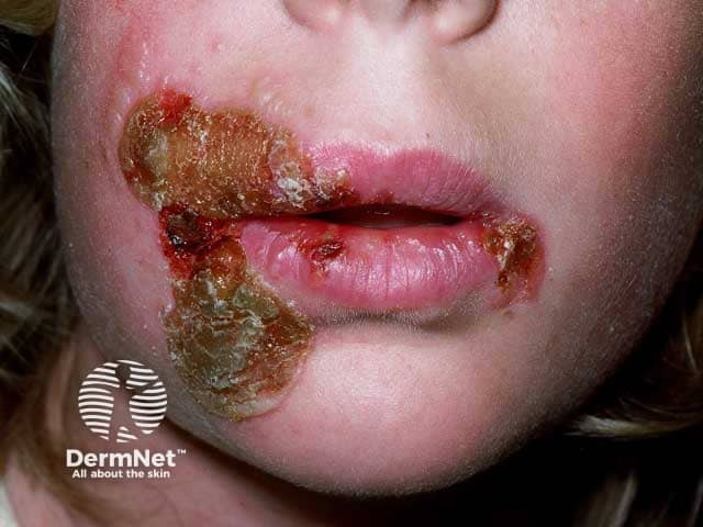
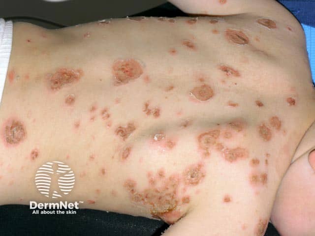
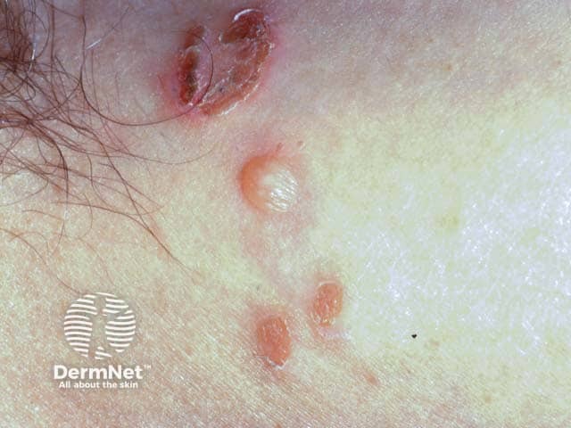
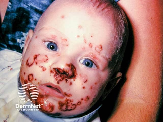
Impetigo is a common, superficial, highly contagious bacterial skin infection characterised by pustules and honey-coloured crusted erosions.
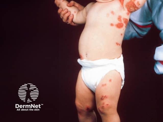
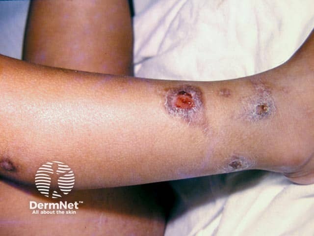
It affects the superficial layers of the epidermis and is typically caused by Staphylococcus aureus and Streptococcus pyogenes (Group A beta – haemolytic streptococci (GABHS)). It can be classified into non-bullous (also known as ‘school sores’) and bullous impetigo. Ecthyma is a deep form of impetigo causing deeper erosions of the skin into the dermis.
Secondary infection of wounds or other skin lesions with the same pathogens is called ‘impetiginisation’.
For more images of impetigo, click here.
Impetigo is most common in young children but can occur at any age. It is usually transmitted through direct contact.
Risk factors which may predispose an individual to impetigo include:
Impetigo is caused by Staphylococcus aureus, and less commonly Streptococcus pyogenes.
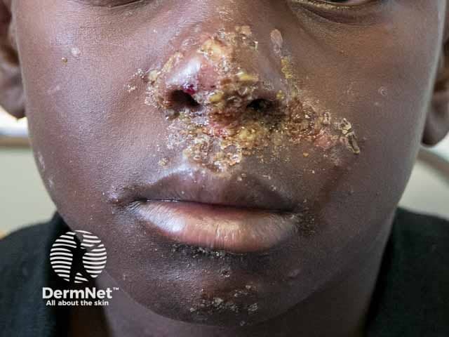
The initial erythematous macule in non-bullous impetigo may be more difficult to see on darker skin tones.
Impetigo is usually self-limiting without serious complications. Without treatment, impetigo usually heals in 2–3 weeks; with treatment lesions resolve within 10 days.
Postinflammatory hypopigmentation or hyperpigmentation may occur but scarring is uncommon.

