DermNet provides Google Translate, a free machine translation service. Note that this may not provide an exact translation in all languages

Translate

Quiz
Leg ulceration – 10 cases

This quiz tests your diagnostic skills for leg ulceration.

For each of the ten cases, study the image(s) and then answer the questions. You can click on the image to view a larger version if required.

Each case should take approximately 2 minutes to complete. There is a list of suggested further reading material at the end of the quiz.

When you finish the quiz, you can download a certificate.
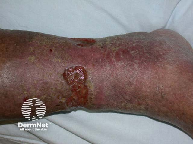
Case 1

A patient presented with chronic ulceration of the lower leg.

Venous ulceration.

Ulcers in association with chronic venous insufficiency usually follow minor traum. Venous disease arises from calf muscle pump dysfunction. It is associated with prior deep venous thrombosis or cellulitis, incompetent (varicose) superficial or communicating veins and immobility.

About 1% of the middle-aged and elderly population is affected by leg ulceration, most commonly in association with chronic venous sufficiency (45-80%).
Venous insufficiency results in aching, swollen lower legs that feel more comfortable when elevated. Signs may include:

  • Varicose veins
  • Chronic lymphoedema
  • Brawny stippled pigmentation
  • Stasis dermatitis (scratched, dry or weeping patches)
  • Atrophie blanche (scarring with prominent tortuous capillaries)
  • Lipodermatosclerosis (firm to hard induration)
  • Warty papillomatosis

A minor injury causes a punched-out ulcer that expands with an irregular brownish border. It may be painless or painful, dry or oozy. The most common site is the medial lower aspect of the calf especially over the malleolus. Multiple ulcers may occur.

Mark question

Sign up to the newsletter