DermNet provides Google Translate, a free machine translation service. Note that this may not provide an exact translation in all languages

Translate

Quiz
Skin coloured lumps and bumps – 12 cases

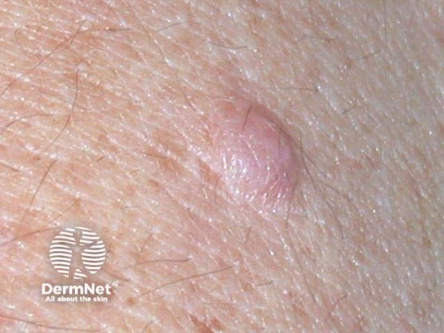
Skin coloured lumps and bumps may be difficult to distinguish one from another. It is helpful to consider body site, location within the skin, size, consistency and morphology. Some common lesions are described here.

First, a reminder about terminology in dermatology.

  • A papule is a small palpable lesion (less than 0.5 cm)
  • A nodule is a larger rounded lesion
  • A plaque is a flat palpable lesion
  • A cyst is fluctuant because it contains fluid or semi-fluid material

For each of the twelve cases, study the image(s) and then answer the questions. You can click on the image to view a larger version if required.

Each case should take approximately 2 minutes to complete. There is a list of suggested further reading material at the end of the quiz.

Case 1

Dermal melanocytic naevus

Moles, or melanocytic naevi, may occur at any site and are quite variable in appearance. They mostly present as stable and well-demarcated papules, nodules and plaques. Moles first appear during childhood, adolescence and early adult life. They vary in colour depending on the activity of melanocytes and the depth of pigment within the skin. Skin coloured moles are dermal naevi without melanin. They may be soft to firm in consistency. They may be pedunculated and ‘wobbly’, or more deeply situated and fixed in position. Histology reveals nests of intradermal melanocytes, which may be neural-type i.e. they may have a spindle-shaped structure.

Mark question

Sign up to the newsletter