

Blue skin lesions – 7 cases
ADVERTISEMENT
Blue skin lesions may be due to melanocytic lesions, vascular lesions or exogenous pigment. Dermoscopy may be useful to distinguish them.
Melanin is normally a brown colour. Melanocytic lesions tend to be blue in colour if the melanin is located within the dermis. They may be congenital (e.g., mongolian spot) or acquired.
For each of the seven cases, study the image(s) and then answer the questions. You can click on the image to view a larger version if required.
Each case should take approximately 2 minutes to complete. There is a list of suggested further reading material at the end of the quiz.
Case 1
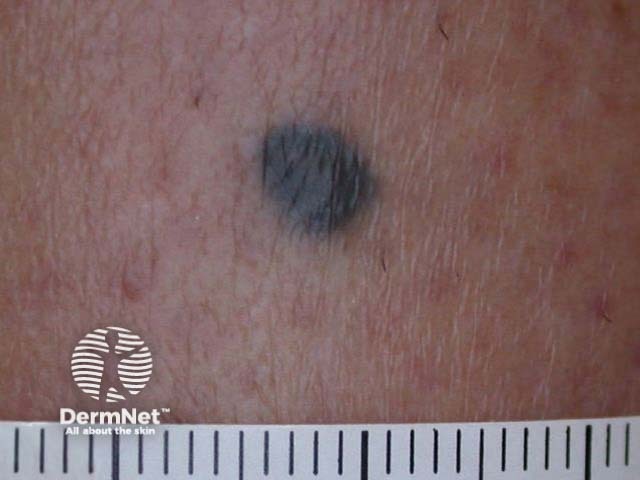
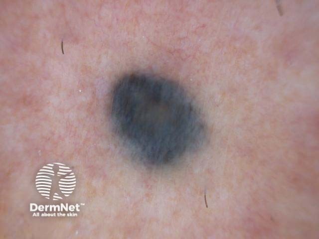
Name this blue-coloured skin lesion.
Describe the dermoscopic features
ADVERTISEMENT