

Oral hairy leukoplakia pathology
Figure 1
Keywords: Histopathology-image, Oral hairy leukoplakia, Pathology
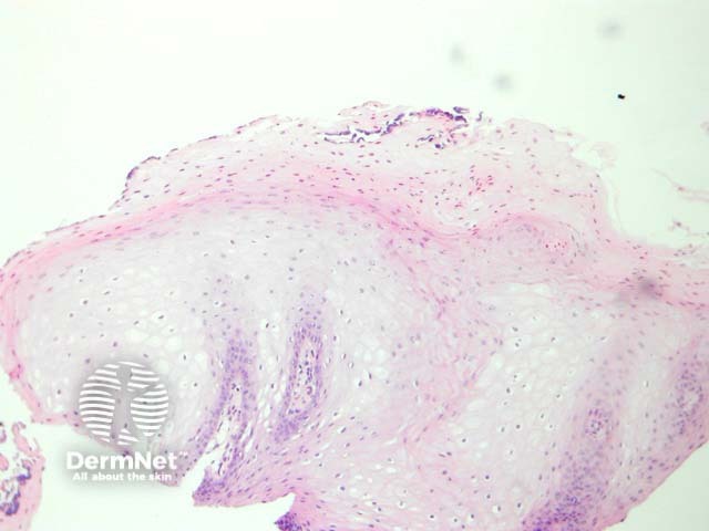
Histology of oral hairy leukoplakia
In oral hairy leukoplakia, the mucosa displays mild papillary acanthosis (figure 1). Hyperkeratosis and marked parakeratosis of the superficial epithelial layer is a prominent feature (figure 2). Superficial infections of the hyperkeratinized epithelium with bacteria or Candida may also be seen. The acanthosis is caused by ballooning koilocyte-like cells. (Figures 1, 3). The nuclei of these have a homogenous ground-glass appearance and may contain intranuclear inclusions.
© DermNet
You can use or share this image if you comply with our image licence. Please provide a link back to this page.
For a high resolution, unwatermarked copy contact us here. Fees apply.
Source: dermnetnz.org