

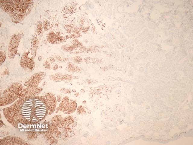
Malignant basomelanocytic tumour pathology
Figure 3
Keywords: Histopathology-image, Malignant basomelanocytic tumour, Pathology
Histology of malignant basomelanocytic tumour
Low power examination of malignant basomelanocytic tumour show an infiltrating dermal tumour with features of a basal cell carcinoma replete with peripheral palisading and retraction around the tumour islands (figure 1). Higher power magnification may be needed to identify malignant melanocytes. Prominent nucleoli can be a helpful feature as these are not typically seen in regular basal cell carcinomas (figure 2).
© DermNet
You can use or share this image if you comply with our image licence. Please provide a link back to this page.
For a high resolution, unwatermarked copy contact us here. Fees apply.
Source: dermnetnz.org