DermNet provides Google Translate, a free machine translation service. Note that this may not provide an exact translation in all languages

Translate

Quiz
Benign melanocytic naevi – 12 cases

This quiz will test your knowledge of benign melanocytic naevi.

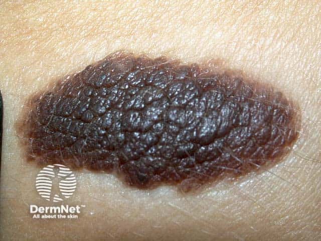
Melanocytic naevi may be congenital or acquired. Melanocytes are normally separated from each other along the basal layer of the epidermis, but in naevi they form nests in contact with each other within the epidermis and/or dermis. They may be found on any cutaneous or mucosal site. Groups of benign naevus cells are sometimes also found in lymph nodes, where they are known as ‘rests’.

For each of the twelve cases, study the image(s) and then answer the questions. You can click on the image to view a larger version if required.

Each case should take approximately 2 minutes to complete. There is a list of suggested further reading material at the end of the quiz.

When you finish the quiz, you can download a certificate.

Case 1

Congenital melanocytic naevus.

Congenital melanocytic naevi (CMN) are considered hamartomas or developmental malformations composed mostly of melanocytes. They often include terminal hair and other elements of skin. They are present at birth or soon after, and may become more prominent over time. They may be flat or elevated, uniform in colour or heterogeneous.
They are quite variable in size and appearance. Clinical types include:

  • Small CMN: less than 1.5 cm diameter
  • Medium sized CMN: 1.5 to 10 cm diameter
  • Giant CMN in other sites: greater than 10 cm diameter
  • Bathing trunk CMN affecting buttocks

Giant melanocytic naevi have a risk of transforming into melanoma in about 5% of patients. The risk is significantly lower in smaller lesions. It is higher in those that cross the spine, with high risk of leptomelanosis. The primary melanoma may arise within the central nervous system.

Mark question

Sign up to the newsletter