

Perineurioma pathology — extra information
Perineurioma is an uncommon benign peripheral nerve sheath tumour composed exclusively of perineural cells.
Histology of perineurioma
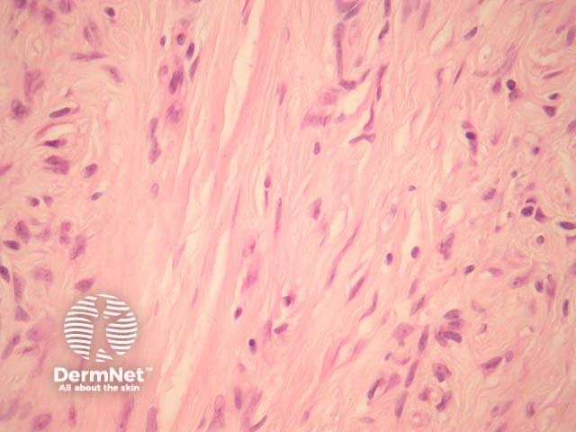
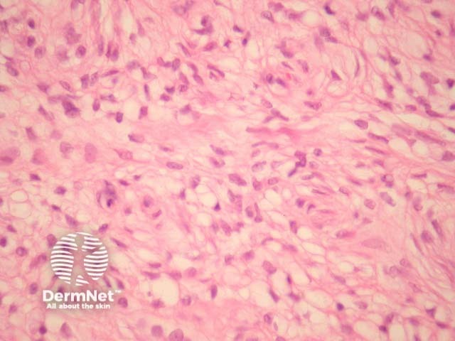
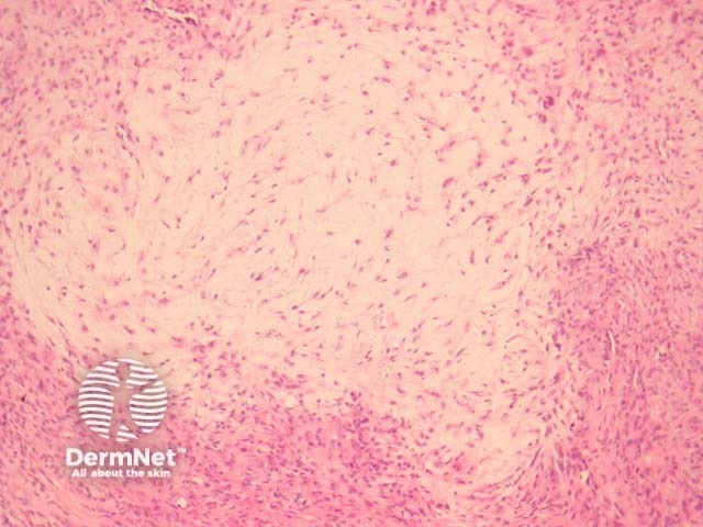
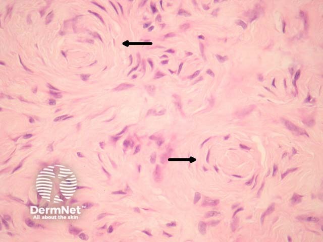
Perineurioma tumour cells are spindle shaped, with long delicate cell processes amidst a collagenous stroma (figure 1). The spindle cells have pale open nuclei with a delicate chromatin pattern, an inconspicuous eosinophilic nucleolus and indistinct cell borders (figures 1, 2). Myxoid change may be seen and can cause diagnostic confusion (figure 3). Whorls of cells are often seen which are reminiscent of meningioma (figure 4, arrows).
Special studies for perineurioma
Immunohistochemical studies show membranous positivity with EMA.
The tumour cells are negative for S100 protein, cytokeratin, desmin and smooth muscle actin.
Differential diagnosis of perineurioma pathology
Dermatofibroma – Typically show polymorphous cell population with more prominent inflammatory cells and often contains multinucleate giant cells. These are EMA negative.
Neurothekeoma – These tumours display a characteristic lobular or nested growth pattern. These are consistently negative with EMA.
Neurofibroma – Neurofibromas are positive with S100 and are composed of other components of peripheral nerves (not just perineural cells).
References
- Weedon’s Skin Pathology (Third edition, 2010). David Weedon
- Robson AM, Calonje E. Cutaneous perineurioma: a poorly recognized tumour often misdiagnosed as epithelioid histiocytoma. Histopathology. 2000 Oct;37(4):332–9. PubMed
On DermNet