

Silver nitrate pigmentation pathology — extra information
Categories:
ICD-11:
SNOMED CT:
ADVERTISEMENT
Silver nitrate can be mistaken for melanoma clinically. It typically presents on patients who work with the agent and unwittingly spill some of the agent onto their skin.
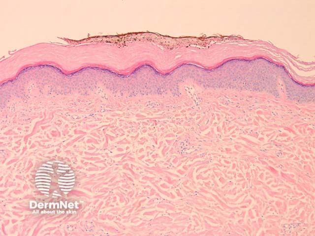
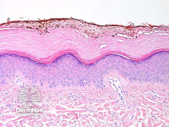
Histology of silver nitrate pigmentation
Brown-black granules are present in the stratum corneum (figures 1, 2). The illustrated case was removed due to a dermatoscopic suspicion of melanoma. The patient was a GP who had recently used silver nitrate for a medical procedure.
Special studies for silver nitrate pigmentation
None are generally needed if the correct history can be elicited.
Differential diagnosis of silver nitrate pigmentation
Melanoma – Careful examination for a melanocytic lesion is always warranted when the clinical impression is suspicious for melanoma
Tinea nigra – Pigmented fungi should be easily distinguished if the morphology is carefully examined
References
- Pathology of the Skin (Fourth edition, 2012). McKee PH, J. Calonje JE, Granter SR
On DermNet
Books about skin diseases
ADVERTISEMENT
Other recommended articles
ADVERTISEMENT