

Synovial metaplasia pathology — extra information
Synovial metaplasia is a change seen most frequently in the tissues surrounding silicone breast prostheses and in healing tissue adjacent to joint prostheses. In the skin and soft tissues, identical features may be seen, frequently in healing or healed traumatic or surgical wounds.
Histology of synovial metaplasia
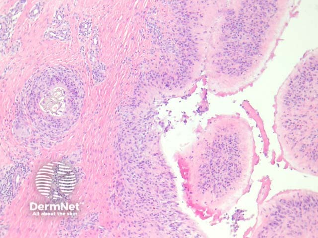
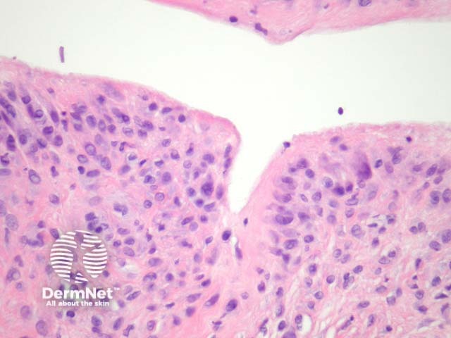
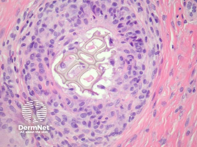
Sections show a irregularly shaped cystic space without an epithelial lining (ie a pseudocyst) surrounded by a dense fibrosis (figure 1). The pseudocyst is lined by a characteristic membrane similar to hyperplastic synovium: the cells of the membrane showed an eosinophilic spindle shaped cytoplasm with processes towards the lumen, which is identical to synovium (figure 2, right hand side; figure 3). It is not uncommon to find foreign material (figure 2, left hand side; figure 4) surrounded by a foreign body reaction and/or synovial metaplasia.

Special studies for synovial metaplasia
None are generally needed
Differential diagnosis of synovial metaplasia
Synovium – Hyperplastic synovium can be impossible to distinguish from synovial metaplasia. Distinction can be impossible in soft tissue adjacent to a joint space. Identification of foreign material favours synovial metaplasia.
References
- Fowler MR, Nathan CO, Abreo F. Synovial metaplasia, a specialized form of repair. Arch Pathol Lab Med. 2002 Jun;126(6):727–30. PubMed