

Syphilis pathology — extra information
Syphilis is an infectious disease caused by the spirochaete Treponema pallidum.
Histology of syphilis
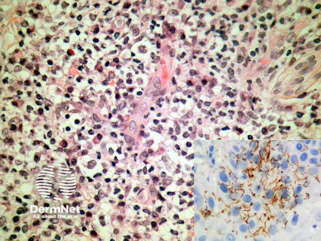
Primary syphilis (primary chancre) demonstrates an acanthotic epidermis which erodes with time to become ulcerated. Under the ulcer bed there is typically a dense lymphocytic response, numerous plasma cells, and endothelial swelling (figure 1). There are typically numerous organisms which may be exhibited with a variety of techniques including immunohistochemistry directed against Treponema pallidum (figure 1, inset).
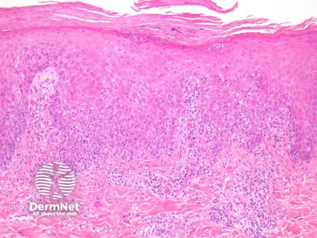
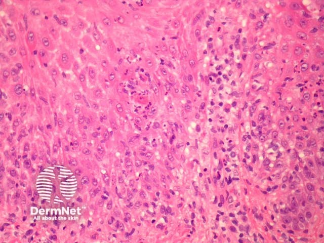
Secondary syphilis exhibits considerable histopathologic variability and may be easily misinterpreted. The epidermis is often involved and shows a psoriasiform hyperplasia with superficial neutrophils (figure 2). There is also a lichenoid tissue reaction, epidermal apoptosis and exocytosis of neutrophils (figure 3). The dermis shows a superficial and deep chronic infiltrate which may resemble the changes of primary syphilis. There are numerous plasma cells in about 1/3 of cases and often endothelial swelling (figure 1).
Tertiary syphilis shows necrotising granulomatous inflammation. The organisms may be impossible to find with special stains and clinical correlation is often needed.
Special studies for syphilis
Various special stains have been applied. The specific immunohistochemical stain for Treponema pallidum is highly specific and sensitive and reveal the organisms to be delicate and spiral shaped (figure 1, inset). Silver impregnation techniques such as Warthin Starry highlight the organisms. Dark-field examination, electron microscopy, and polymerase chain reaction (PCR)-based methods have also been used.
Differential diagnosis of syphilis pathology
Akin to the clinical presentation, the histopathologic presentations of syphilis are diverse. Eczematous, granulomatous, pustular, folliculitis-like, and acantholytic patterns have been described. Clinical correlation and correlation with serologic studies may be useful in addition to the special stains. The presence of plasma cells in the dermis associated with endothelial swelling may point to the diagnosis, particularly when accompanying a psoriasiform epidermal reaction.
References
- Weedon’s Skin Pathology (Third edition, 2010). David Weedon
- Pathology of the Skin (Fourth edition, 2012). McKee PH, J. Calonje JE, Granter SR