

Inflammatory reactions
Created 2009.
Learning objectives
- Recognise inflammatory reactions to systemic disease
Introduction
The dermatoses described on this page are the result of an immunological reaction to underlying systemic disease. In each case, a number of different conditions may be responsible. The severity of the reaction does not necessarily correllate with the severity of the underlying disease.
The mechanisms for the reactions are poorly understood.
Panniculitis
Panniculitis is the term used for a group of disorders in which inflammation primarily affects subcutaneous tissue.
- Erythematous or violaceous nodules in subcutaneous fat
- Nodules may be soft or hard
- May ulcerate or discharge pus or liquefied fat
- Classified according to histology: septal or lobular inflammation, with or without vasculitis
- Idiopathic lobular panniculitis may involve visceral fat or internal organs resulting in fever, nausea and vomiting, abdominal pain and sometimes, death.
- Associated with emphysema in alpha-1-antitrypsin deficiency
- Pancreatitis or pancreatic carcinoma may cause painful liquefying panniculitis
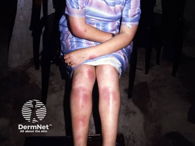
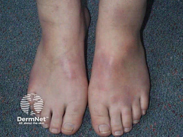
Erythema nodosum
- Subacute septal panniculitis resulting in painful red nodules, mainly of lower legs
- Mostly seen in young adult females
- Often accompanied by fever, malaise and arthralgia especially of ankles
- Resolves in 6 – 12 weeks, depending on aetiology
- Treated by elevating and resting the limbs, salicylates and NSAIDs
- Aetiology includes sarcoidosis, beta haemolytic streptococcal infection, tuberculosis, many other infectious agents, inflammatory bowel disease, malignancies, Behçet syndrome, Sweet disease and drugs (sulphonamides, oral contraceptive pill).

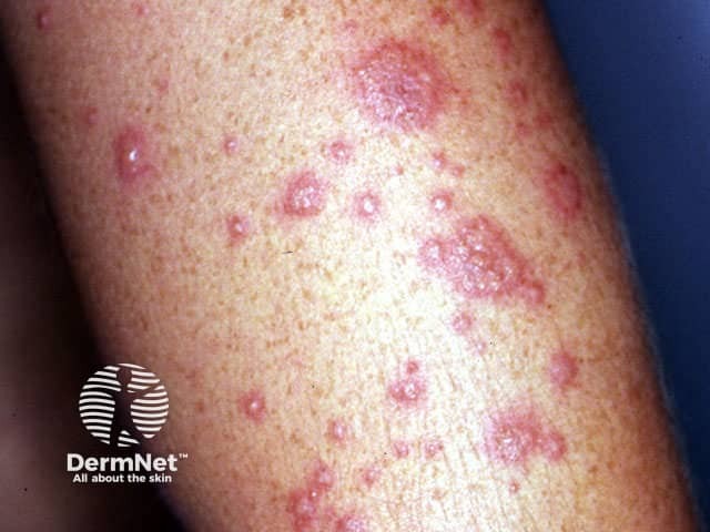
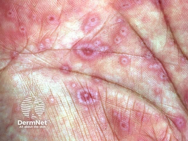
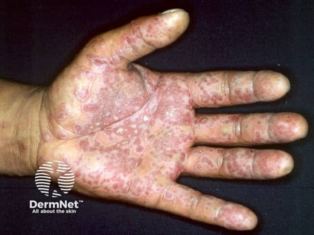
Erythema multiforme
Erythema multiforme (EM) is an acute eruption of erythematous target or iris-shaped plaques that may blister, typically involving the extremities (face, palms, soles and distal limbs).
EM minor
- EM is more common in males than feamles. About 50% are under 20 years of age.
- EM minor is an eruption of classic target lesions on the extremities and persists for one to three weeks associated with mild fever and malaise.
- EM minor is often preceded by infection (herpes simplex, orf, mycoplasma, vaccination).
- Recurrent EM is nearly always due to recurrent herpes simplex and can be prevented by prophylactic oral aciclovir.
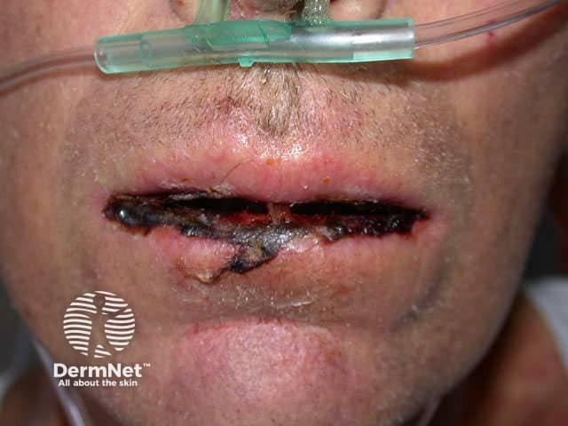
EM major
- EM major or Stevens Johnson syndrome is predominantly a mucosal eruption of erosions and blisters in the oropharynx, on the lips, conjunctivae and genitalia accompanied by fever and prostration.
- Often requires hospitalisation for supportive care.
- EM major is usually a drug eruption especially to sulphonamides, anticonvulsants, allopurinol and antibiotics.
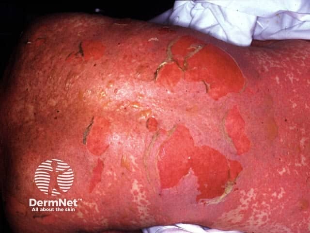
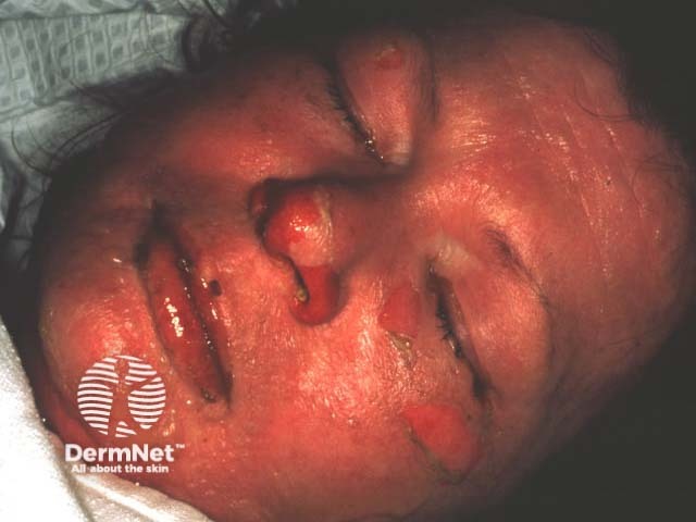
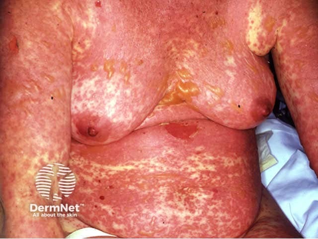
Toxic epidermal necrolysis
- Toxic epidermal necrolysis (TEN) is characterised by widespread tender erythema of skin and mucosa followed by extensive cutaneous and mucosal necrosis and denudation within 2 or 3 days.
- Drugs are responsible for more than 80% of cases of TEN, most often sulphonamides, phenytoin, carbamazepine, allopurinol, lamotrigine and anti-inflammatories, but many drugs have been occasionally implicated. The most recently prescribed medications should be immediately discontinued and should never be readministered.
- Acute graft versus host disease may also result in TEN and is usually fatal.
- TEN is potentially life threatening due to multisystem involvement including renal tubular necrosis, eroded gastrointestinal and respiratory tracts, neutropaenia
- Mechanism related to cell-mediated cytotoxic reaction against epidermal cells.
- Management should be in an intensive care unit and may require early intravenous immunoglobulins or ciclosporin (not corticosteroids) as well as expert supportive care.
Annular erythemas
Several patterns of annular erythema are described, sometimes specific to the underlying aetiology.
- Erythema chronicum migrans: Lyme disease
- Erythema marginatum: acute rheumatic fever
- Erythema gyratum repens: malignancy
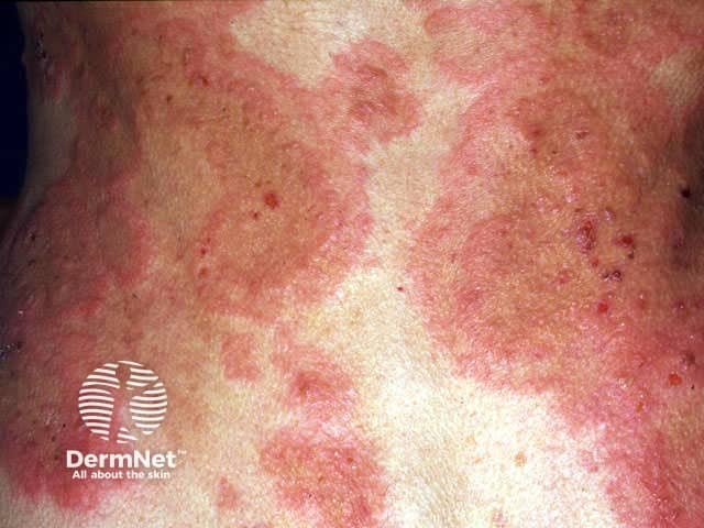
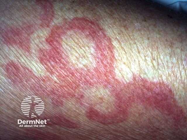
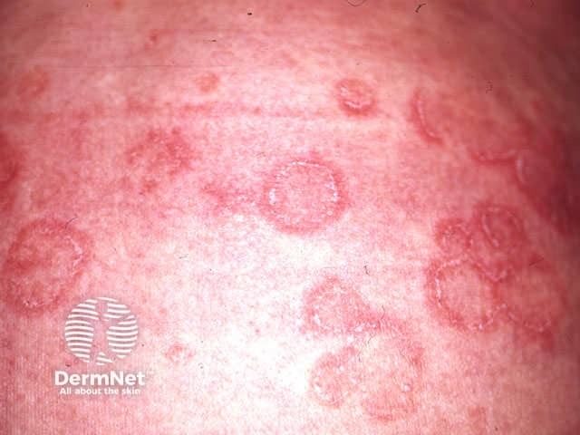
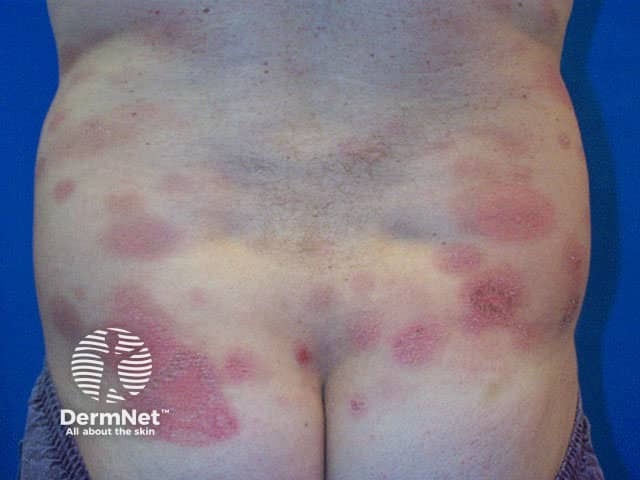
Erythema annulare centrifugum results in expanding erythematous annular and polycyclic plaques, generally on the trunk.
- It may be mildly itchy, and scaly on the trailing edge of the ring.
- It can be confused with many other skin conditions including fungal infection, psoriasis, pityriasis rosea and seborrhoeic dermatitis.
- Underlying causes include dermatophyte infection, candidiasis, drugs and cancer but most cases appear to be idiopathic and chronic.
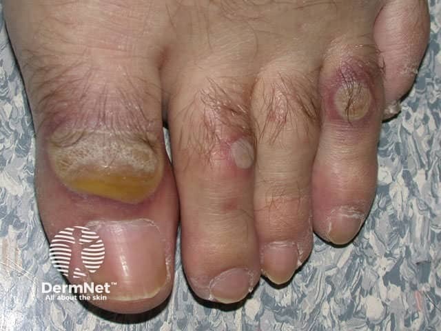
Neutrophilic dermatoses
Dermatoses with dense dermal infiltrates of neutrophils include:
- Pyoderma gangrenosum (PG)
- Sweet disease
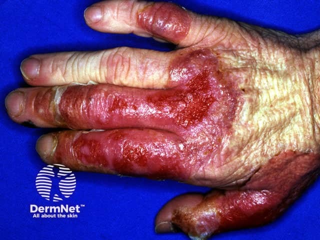
- Neutrophilic dermatosis of the dorsal hands
- Overlap disorders including acute pustular vasculitis
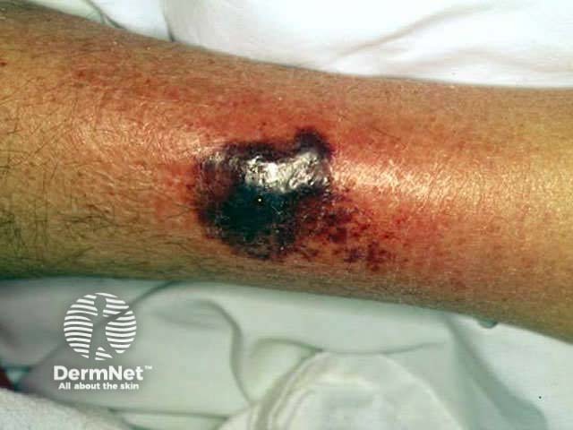
Pyoderma gangrenosum is associated with inflammatory bowel disease, rheumatoid arthritis and haematological malignancies but occasionally arises in otherwise healthy individuals. Sweet disease can have similar causes but more often follows an upper respiratory tract infection.
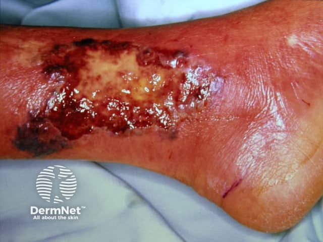
Pyoderma gangrenosum results in severe ulceration characterised by an overhanging necrotic edge and severe pain. It most commonly arises on the lower legs and may be very difficult to control requiring one or more systemic immune suppressive medications.
Pyoderma gangrenosum
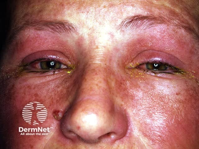
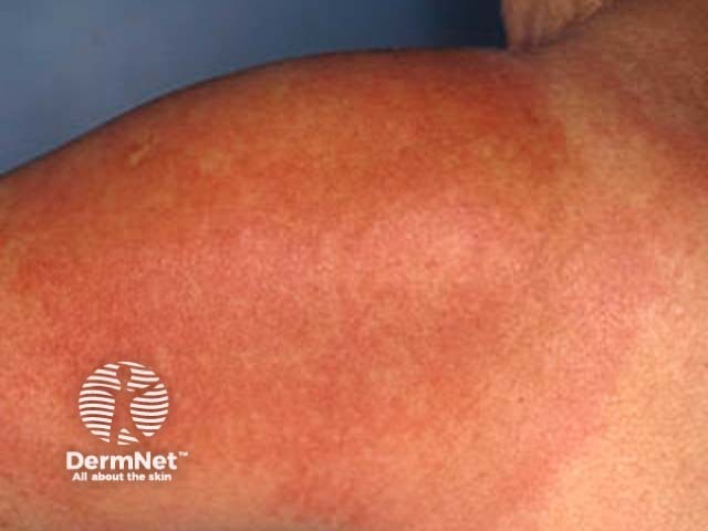
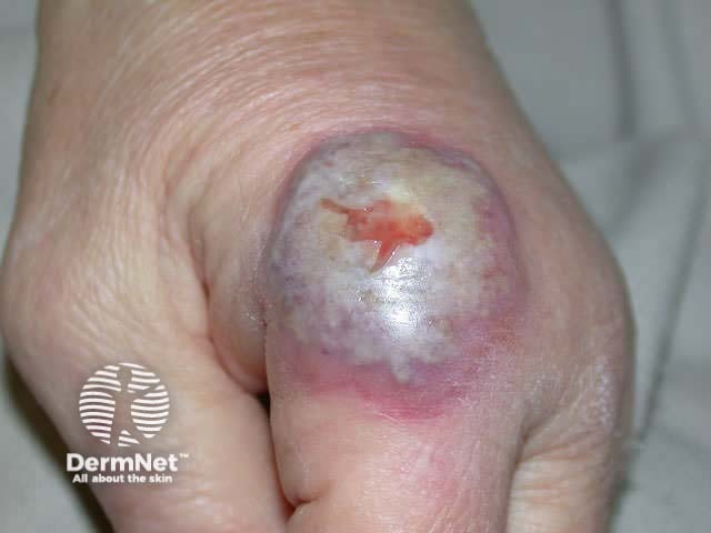
Sweet disease presents as rapidly evolving juicy “pseudo-vesicular” plaques accompanied by fever, leukocytosis, conjunctivitis and arthralgia. The lesions are most often located asymmetrically on the upper extremities, neck and face (cape distribution). They heal within two or three months without scarring, but resolve very quickly if treated with oral prednisone for 4 to 6 weeks.
Sweet disease
Activity
What is the evidence that systemic corticosteroids should be avoided in the management of Stevens Johnson syndrome and toxic epidermal necrolysis?
References:
On DermNet:
Information for patients
Other websites:
- Medscape Reference: Reactive and inflammatory dermatoses
Books about skin diseases:
See the DermNet bookstore