> Go to the image library
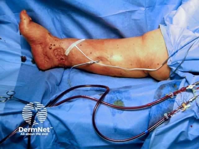
Oedema and wound infection
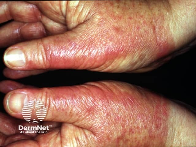
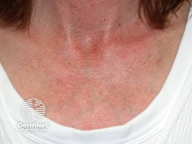
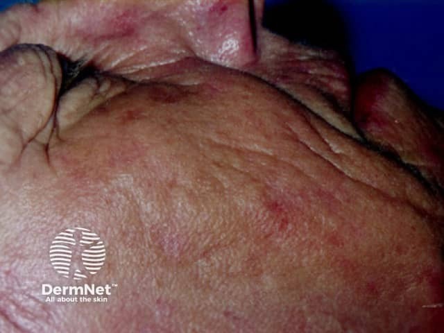
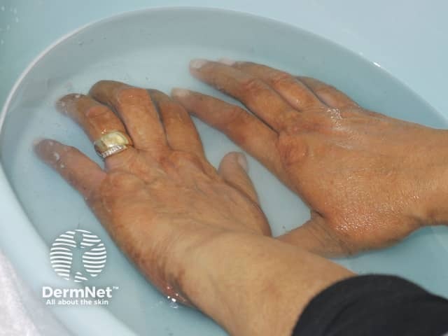
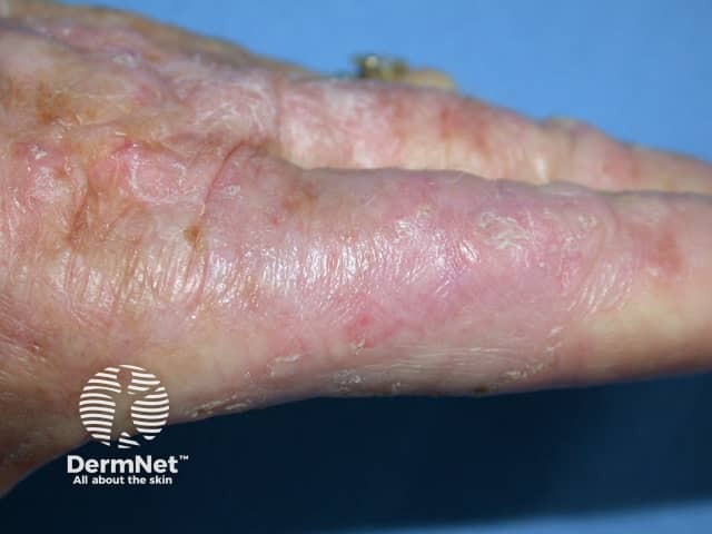
Hands burning after PUVA soaks
Allergy to collagen injected
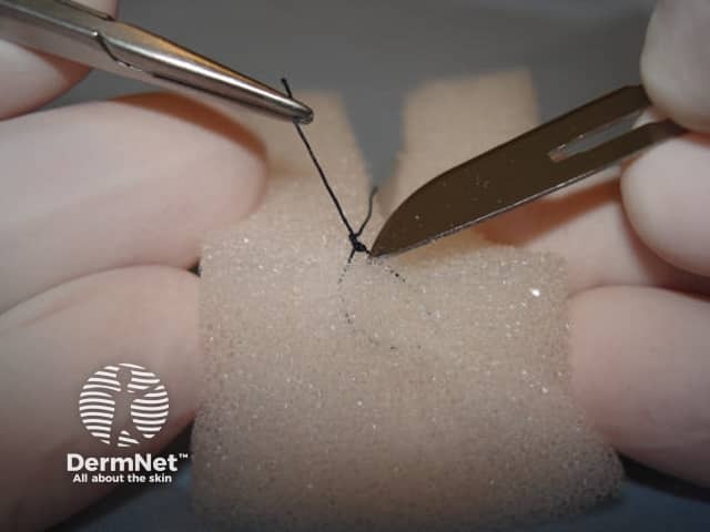
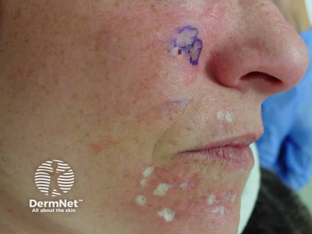
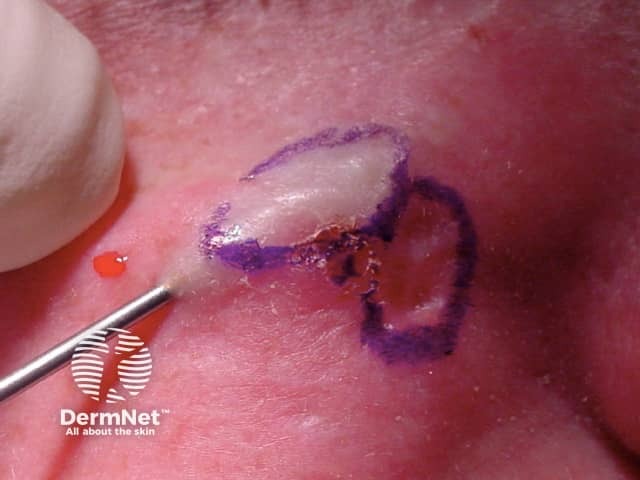
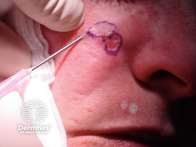
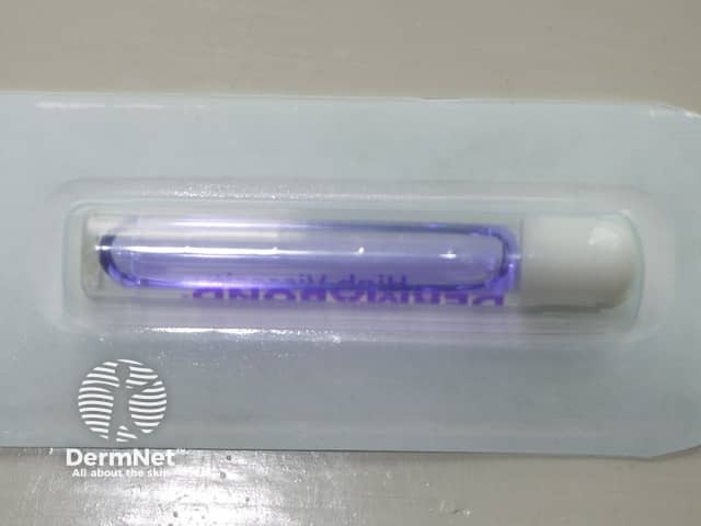
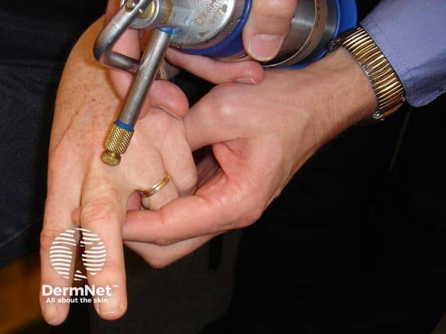
Applying trichloracetic acid for TCA CROSS procedures
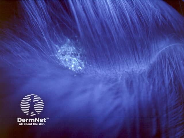
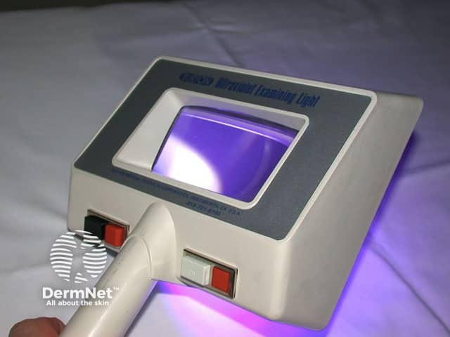
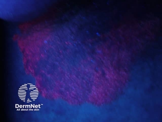
Fluorescence in tinea capitis using Wood lamp
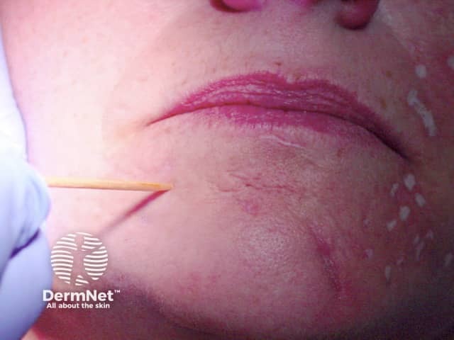
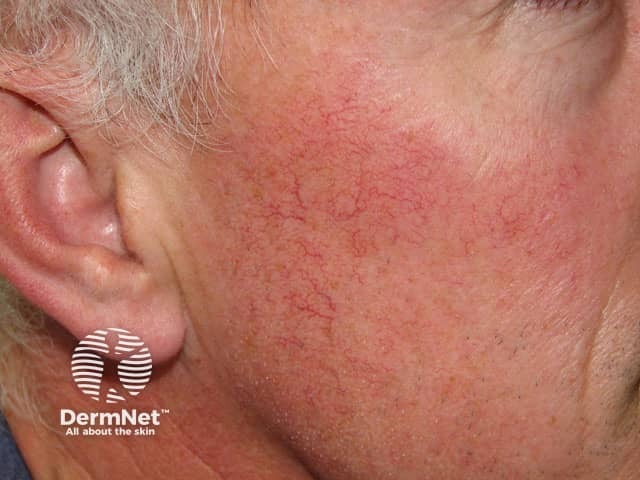
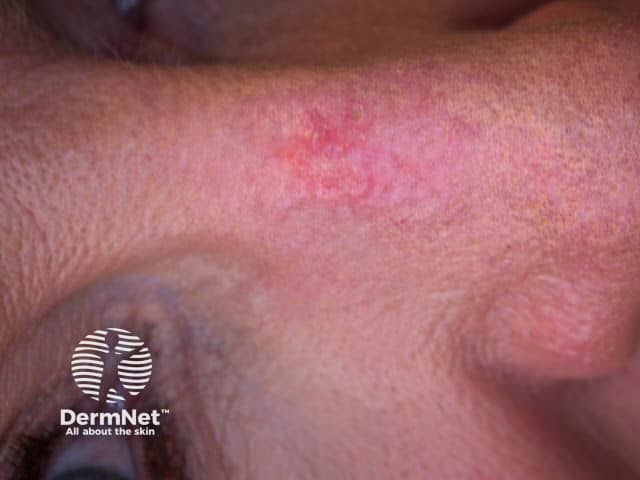
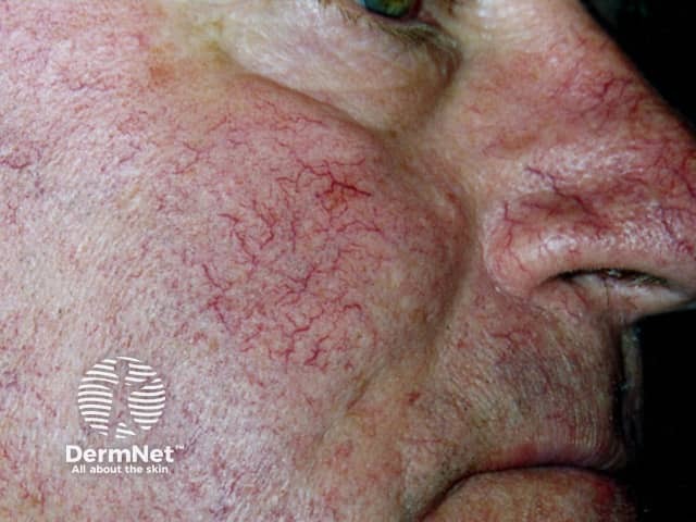
Pulse dye laser for telangiectasia, before treatment
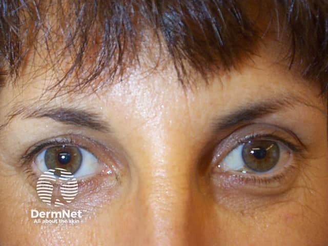
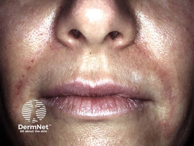
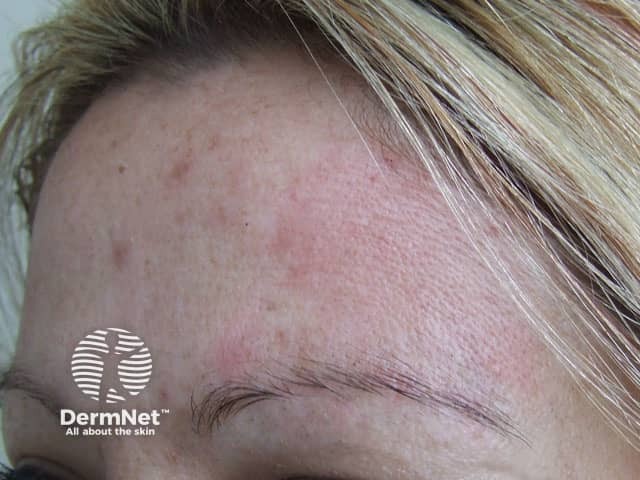
Subject attempting to frown after botulinum toxin injections
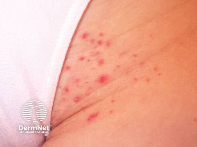
Folliculitis after waxing
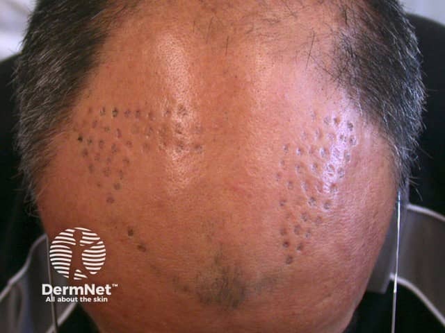
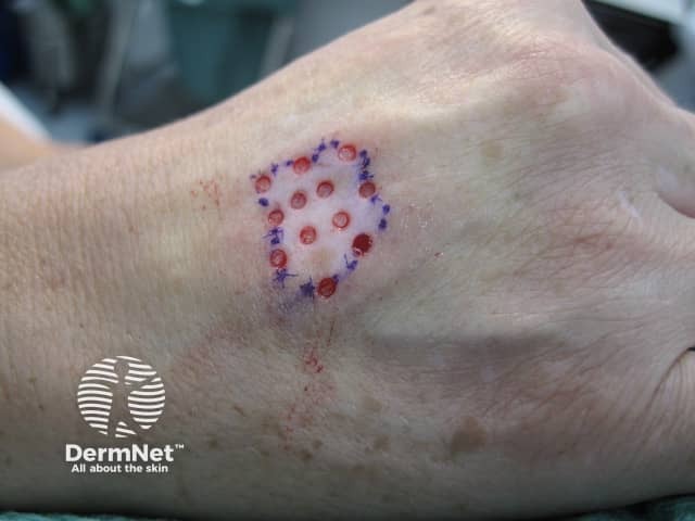
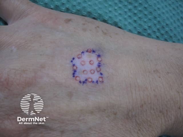
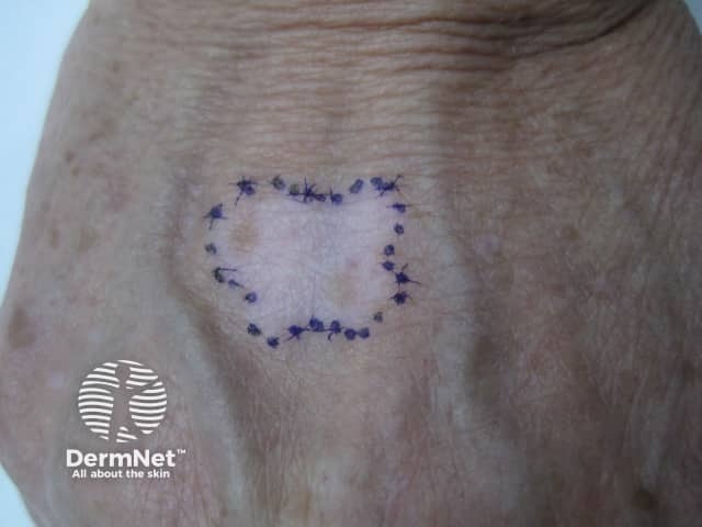
Sentinel node biopsy persistent tattoo
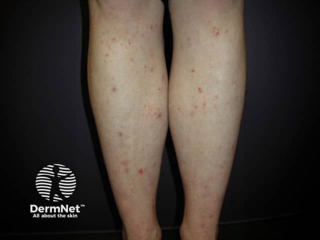
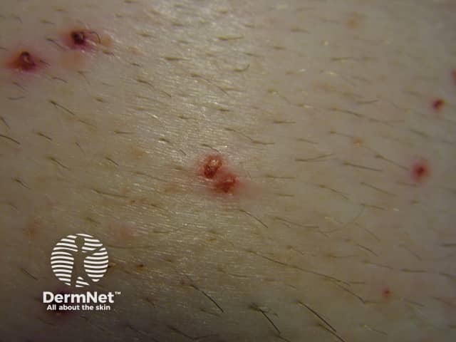
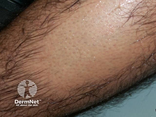
Folliculitis after shaving legs
Hands burning after PUVA soaks
Redness and oedema 4 hrs after cryotherapy
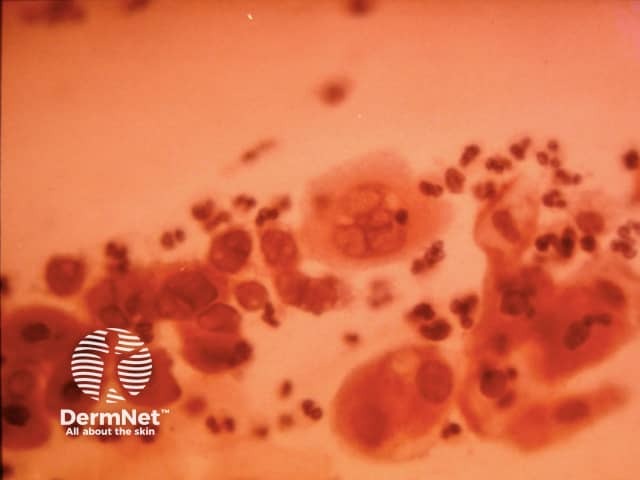
Tzanck smear of herpes simplex
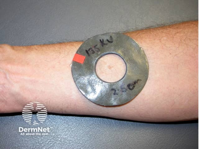
A lead shield is used to protect normal skin and aim the radiation at the target area
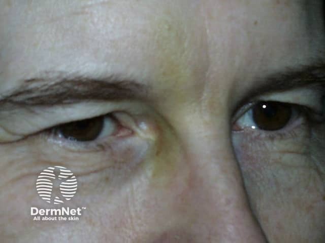
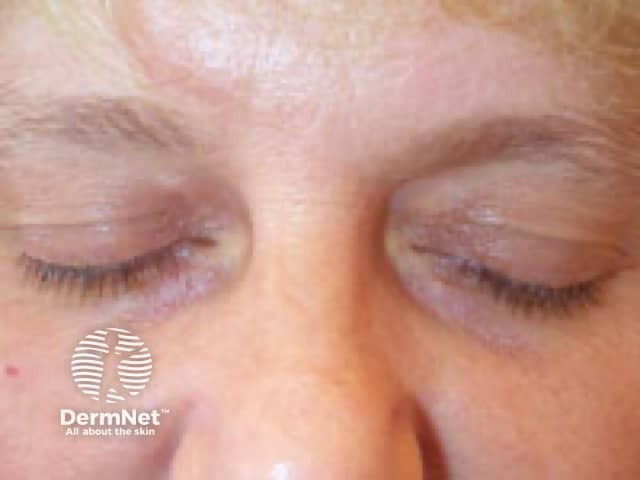
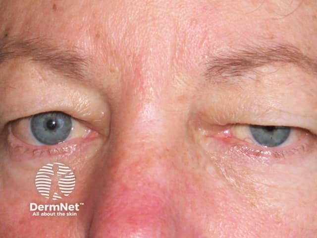
Subject relaxed prior to botulinum toxin injections
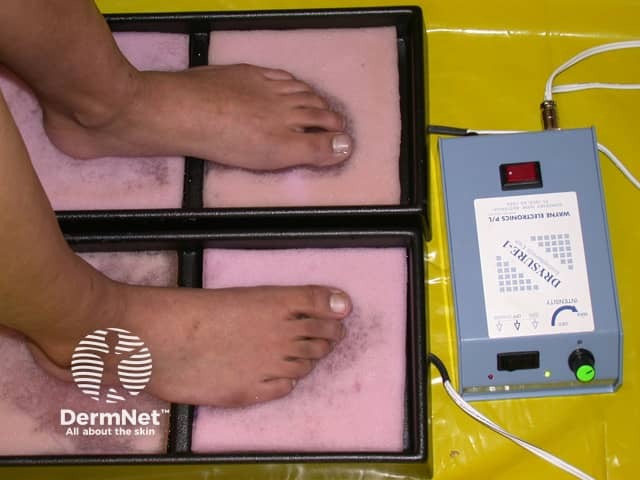
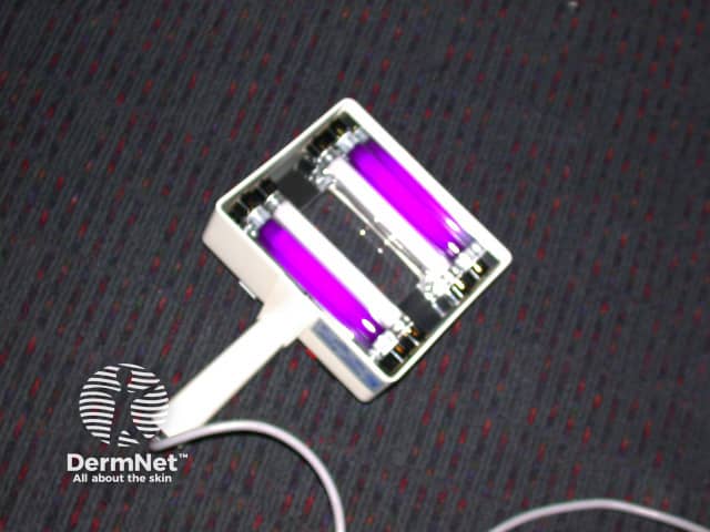
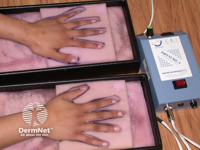
Iontophoresis for plantar hyperhidrosis
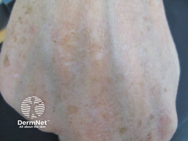
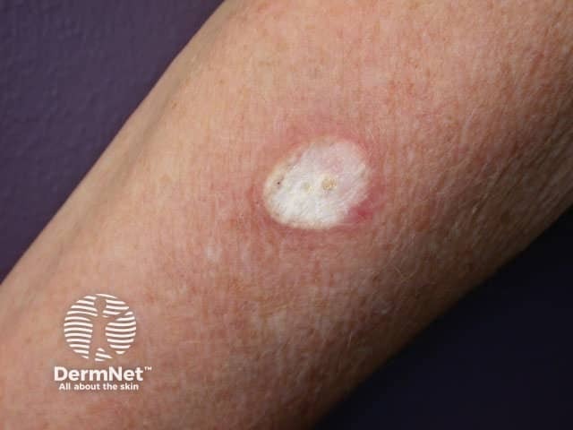
Hypopigmentation following IPL
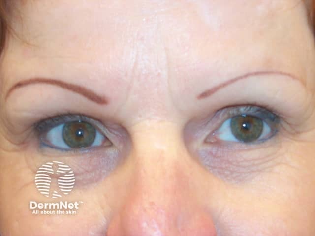
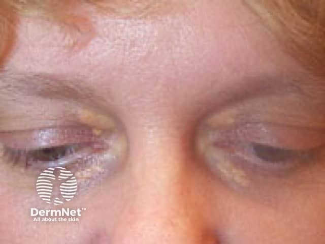
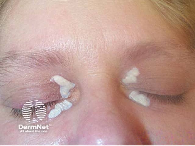
Tattooing (left eyebrow) and additional pencil (right eyebrow)
Hypopigmentation following IPL
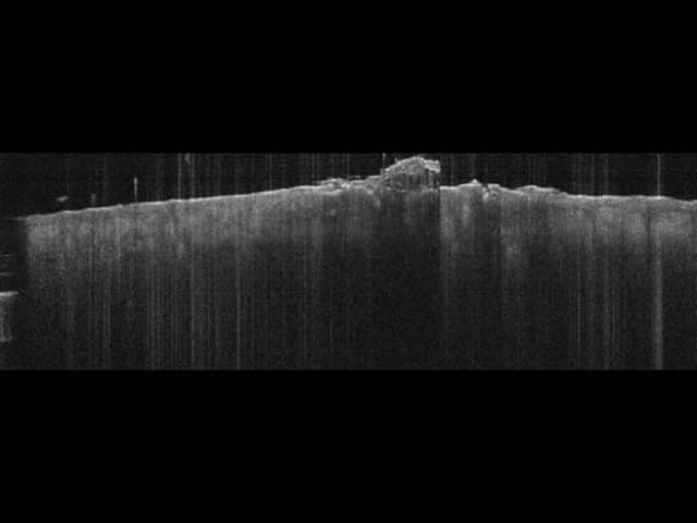
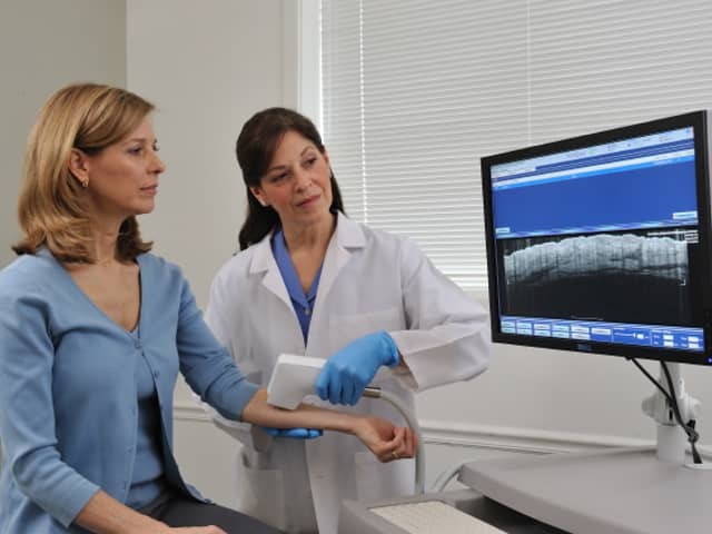
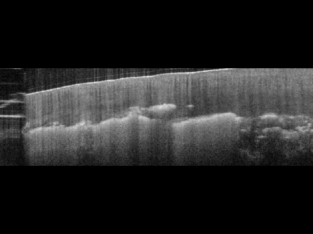
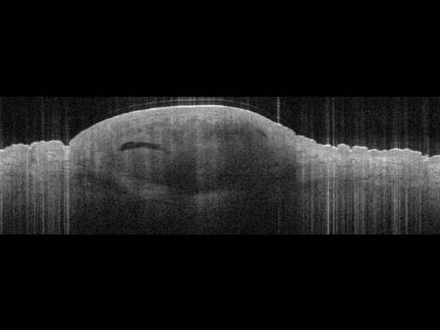
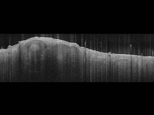
Vertical shadowing from surface scaling (OCT), same lesion
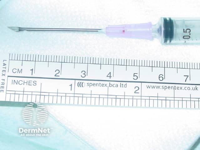
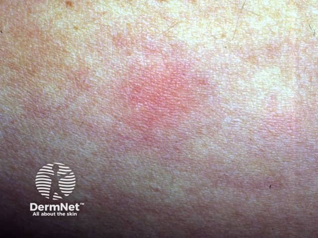
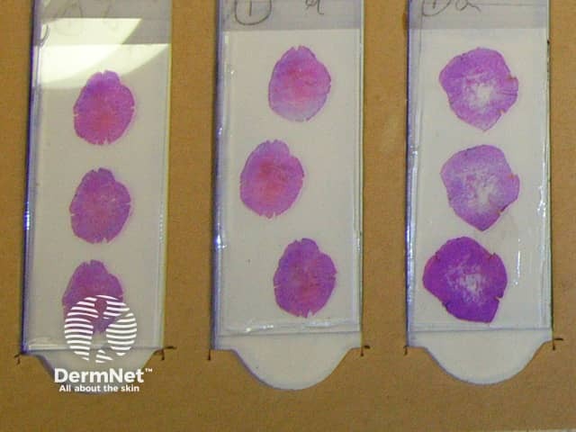
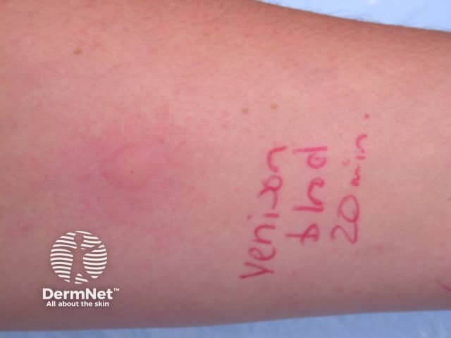
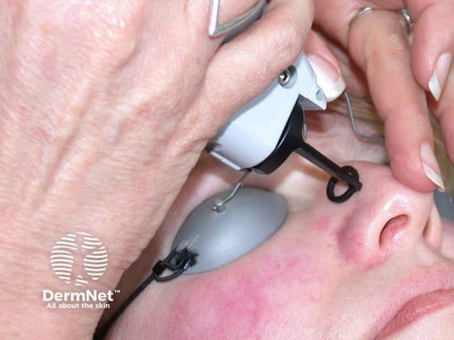
A positive prick test wheal at 20 minutes
Pulse dye laser for telangiectasia, day after treatment
The ice ball just as the cryotherapy is discontinued – it will thaw in 15-30 seconds
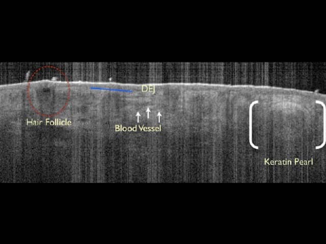
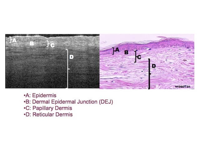
Epidermal and dermal structures
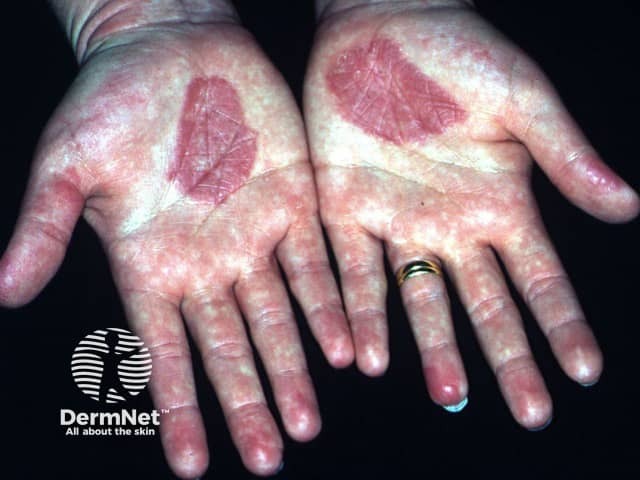
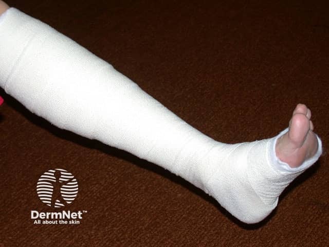
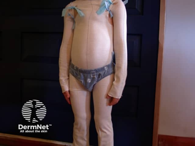
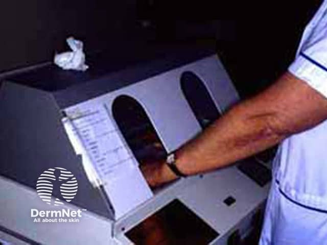
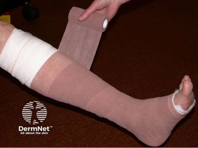
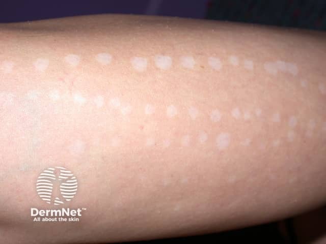
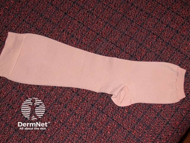
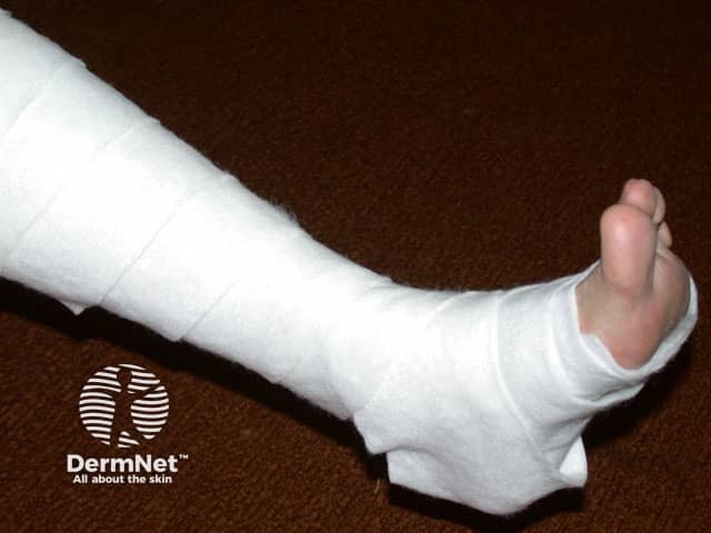
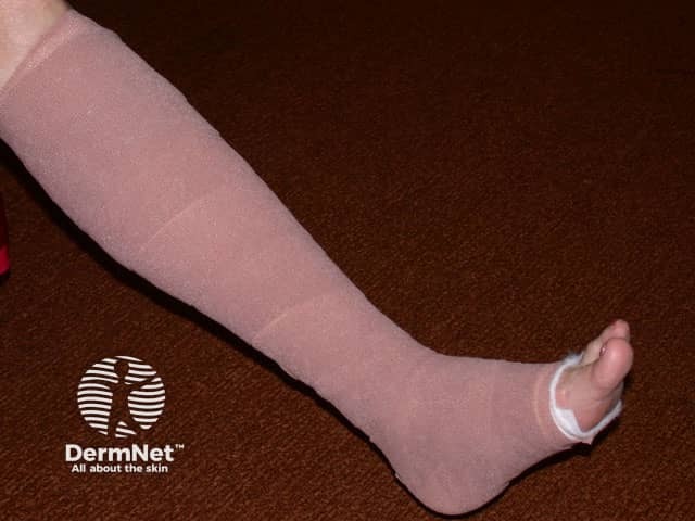
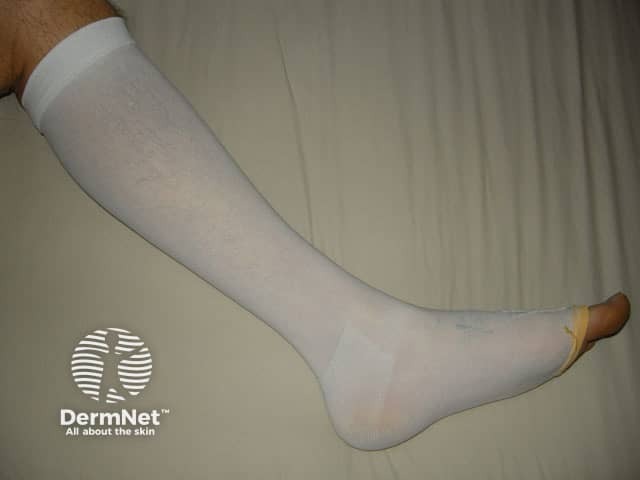
Hands and feet PUVA soaks
Hands and feet PUVA soaks
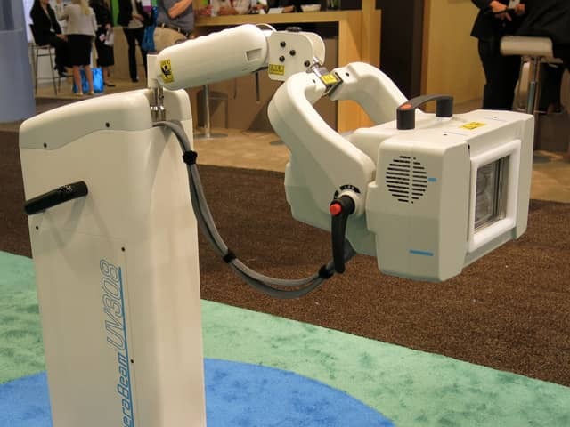
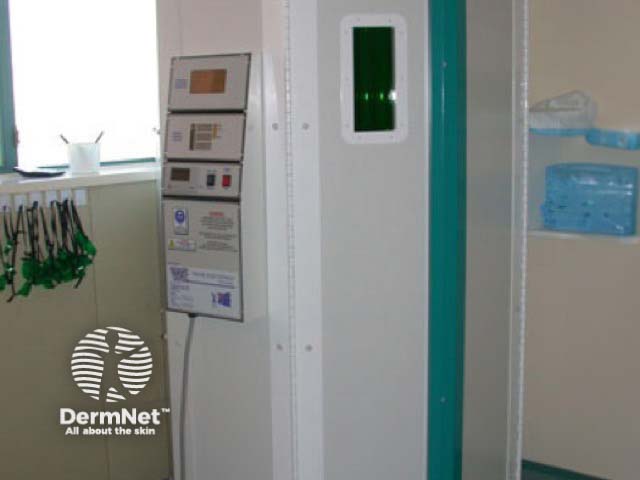
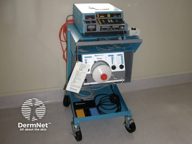
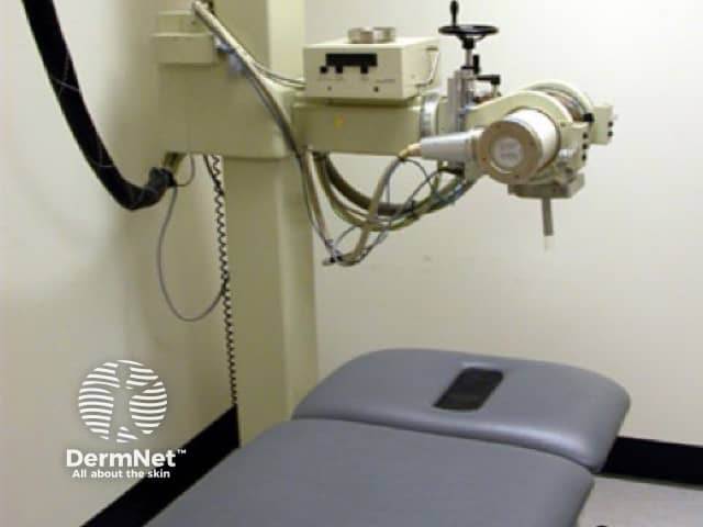
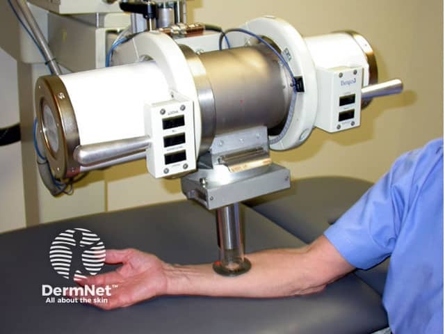
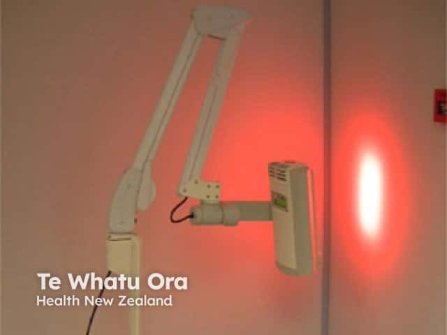
A superficial x-ray machine
Hands and feet PUVA soaks
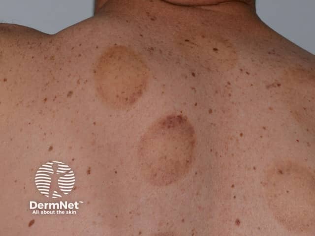
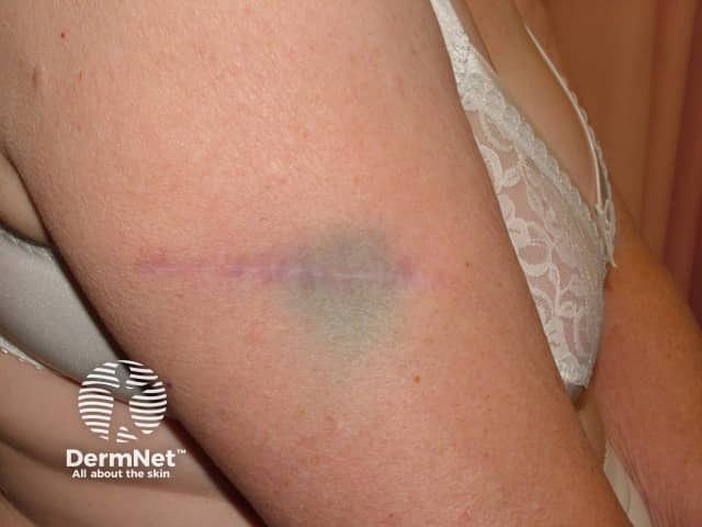
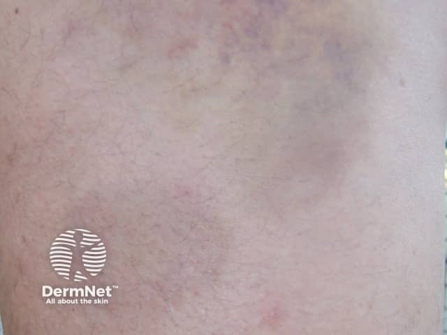
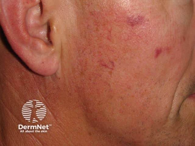
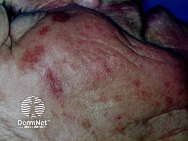
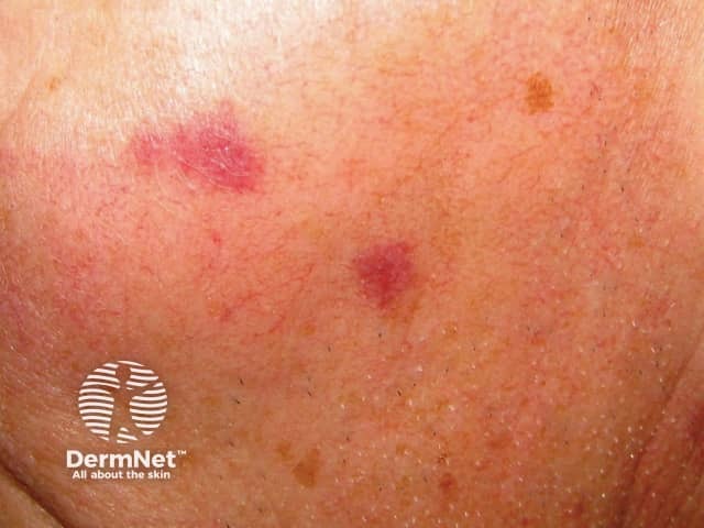
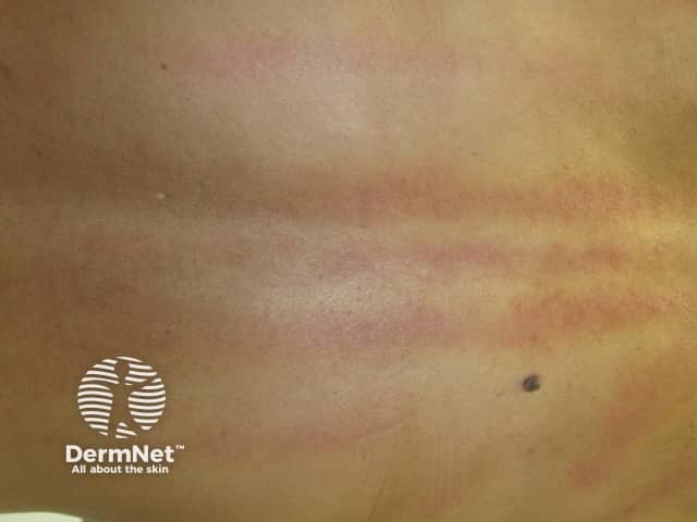
Bruises day after treatment
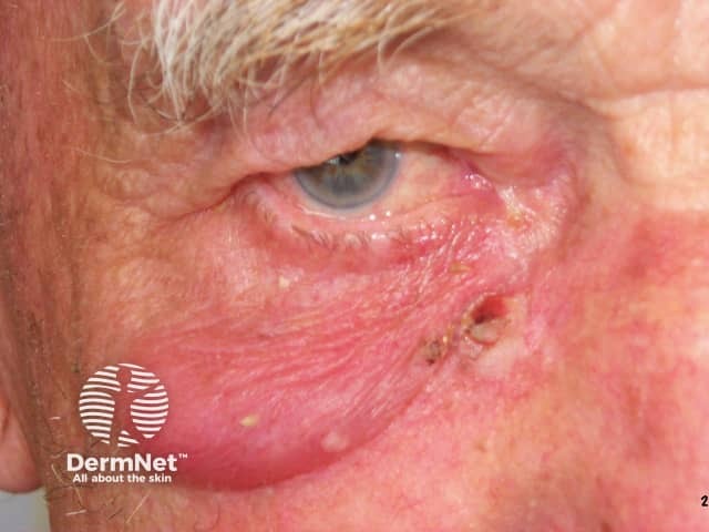
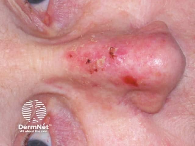
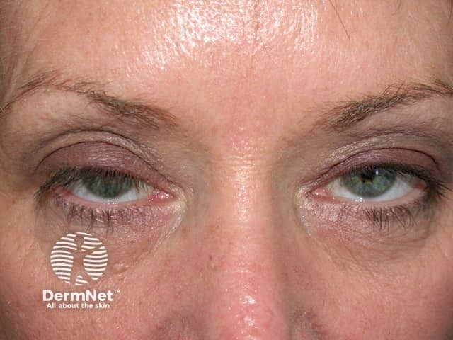
Eyelid oedema 24 hrs after cryotherapy to the lesion adjacent to the nose
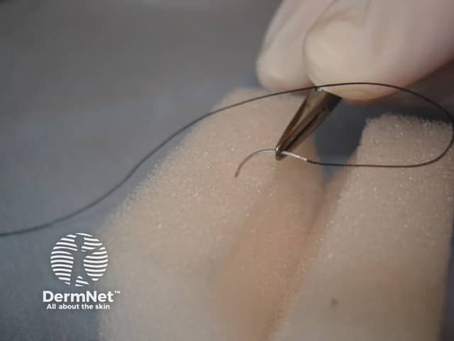
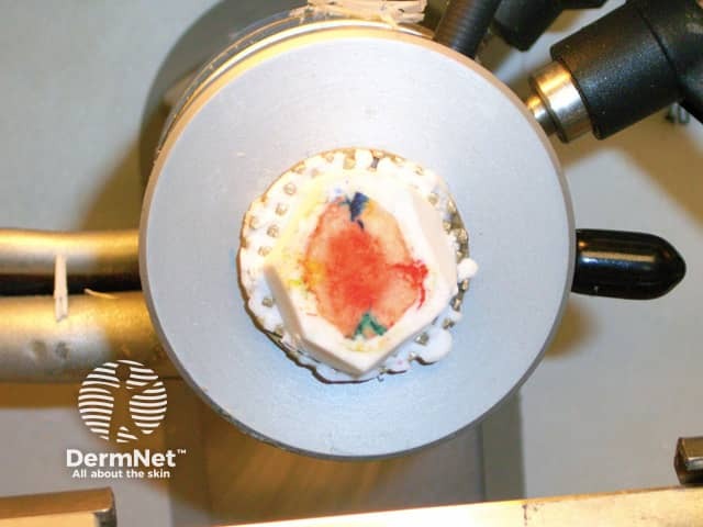
Trichloracetic acid for TCA CROSS procedures
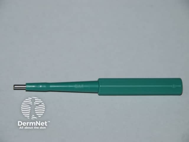
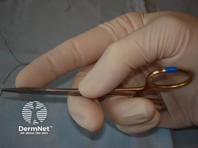
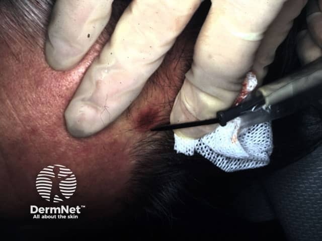
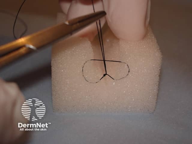
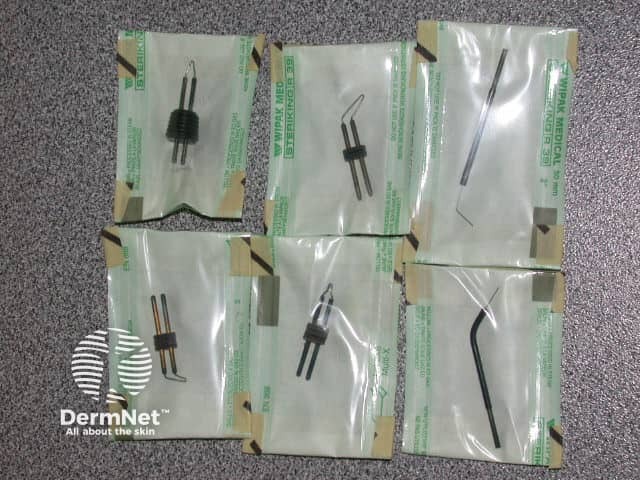
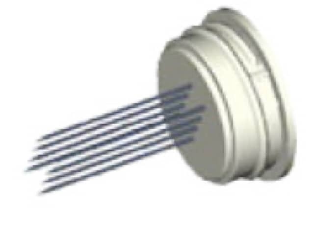
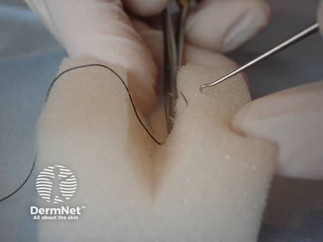
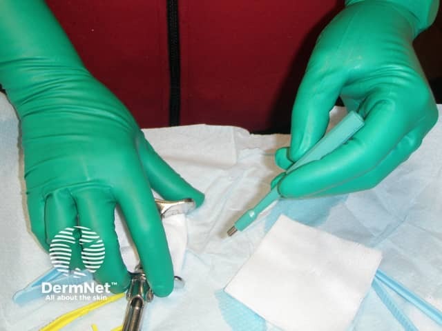
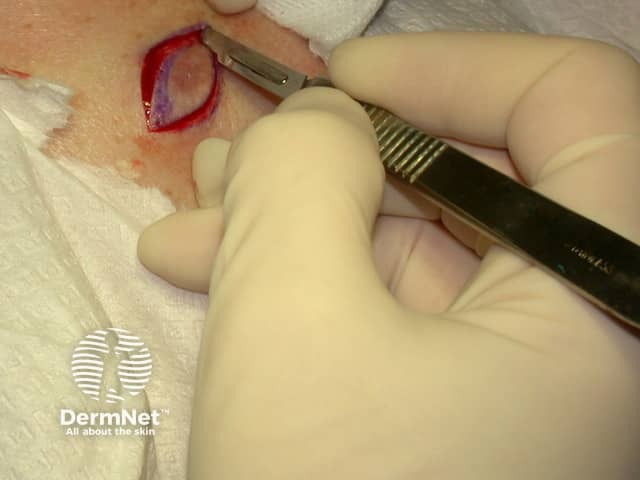
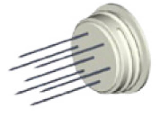
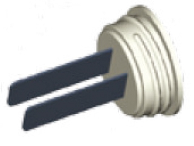
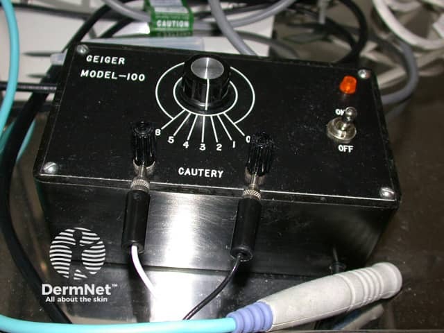
Tips used for electrocautery
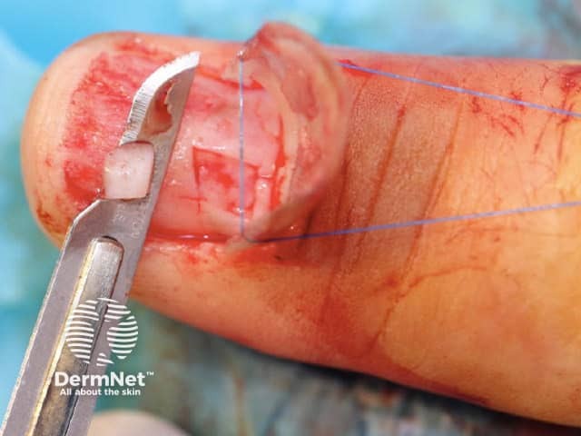
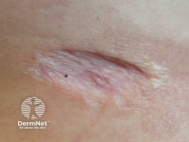
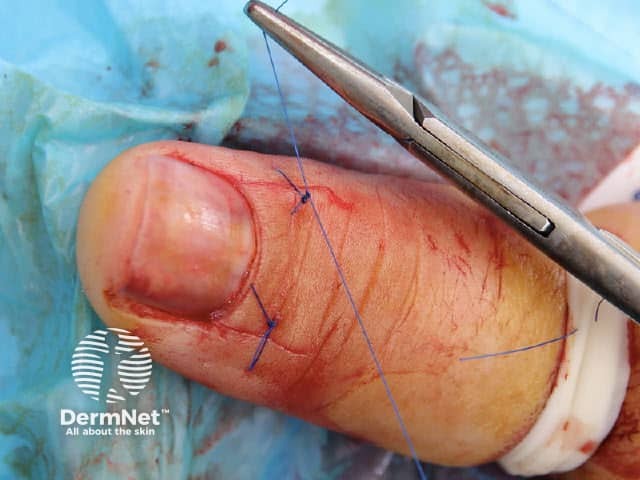
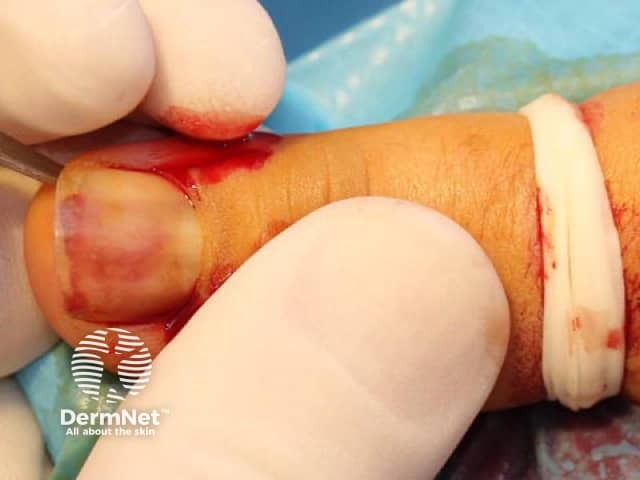
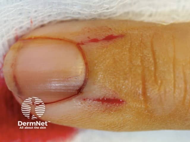
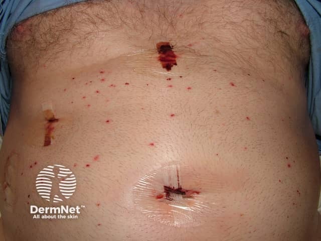
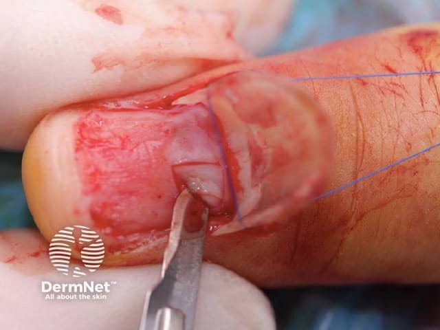
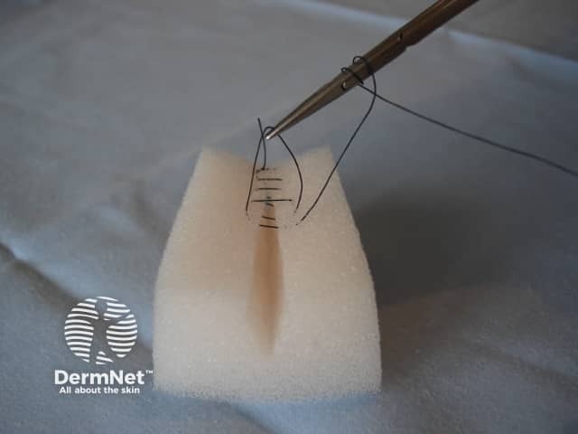
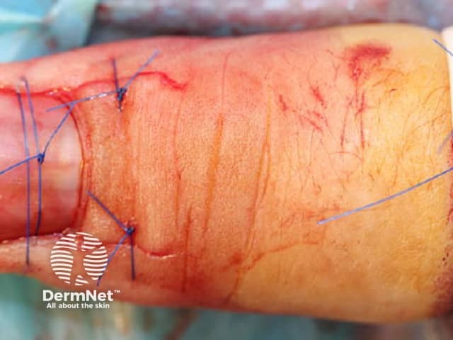
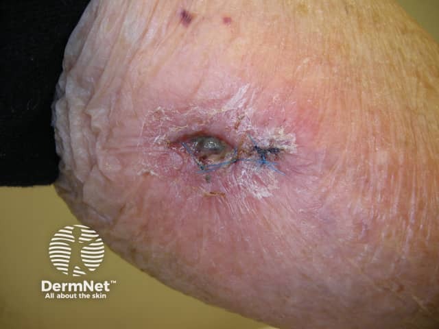
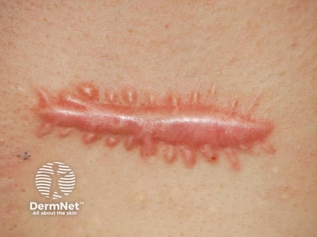
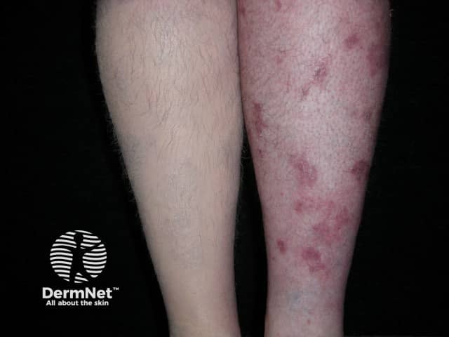
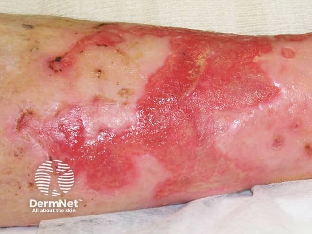
Surgical wound dehiscence
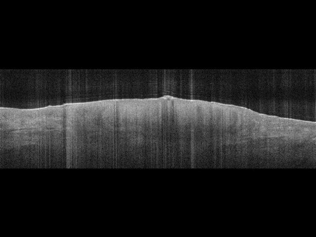
Epidermal and dermal layers on OCT (left) and histopathology (right)
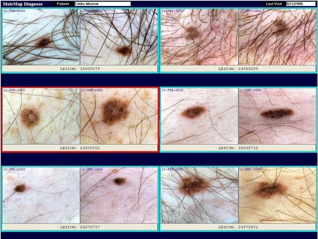
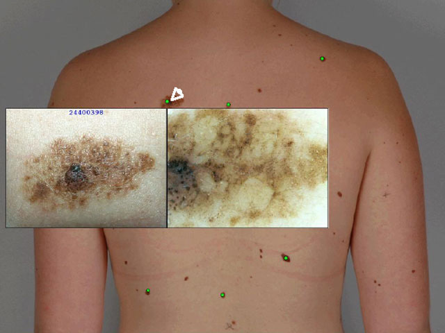
Repeat imaging. Red border indicates lesion has changed
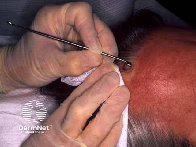
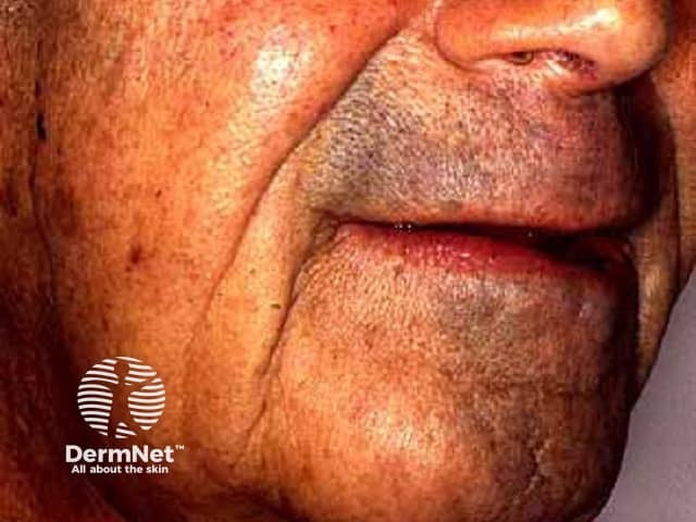
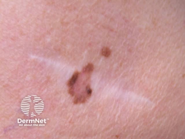
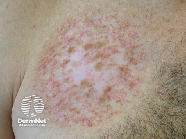
Recurring lentigo maligna
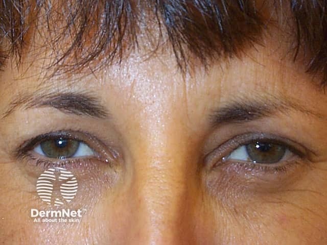
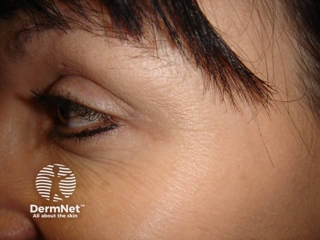
Subject relaxed after botulinum toxin injections
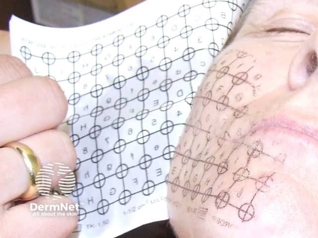
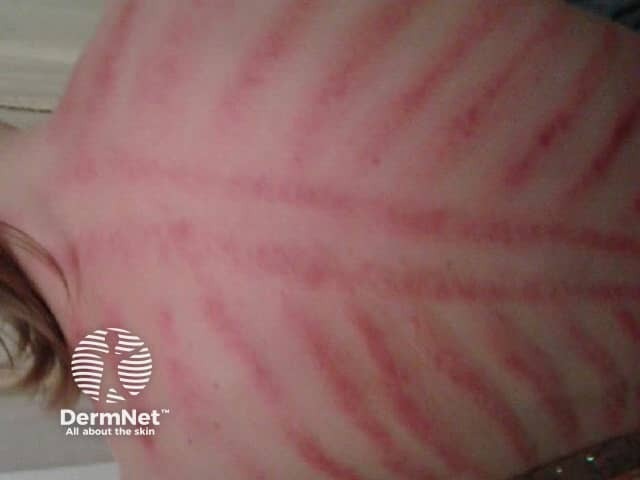
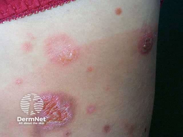
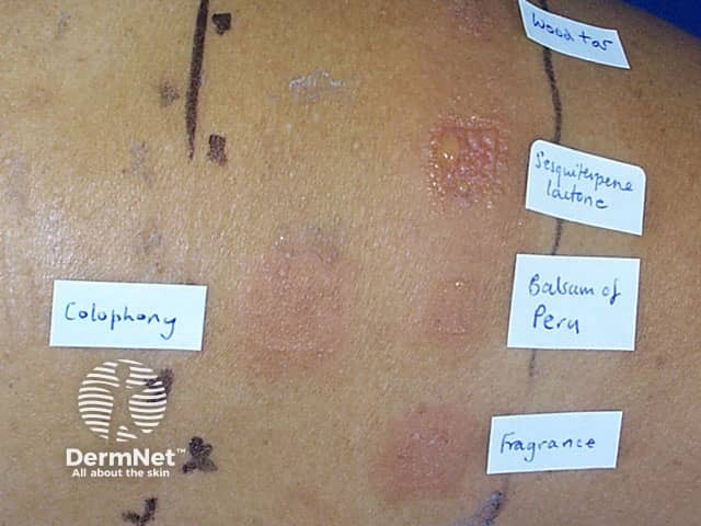
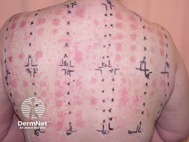
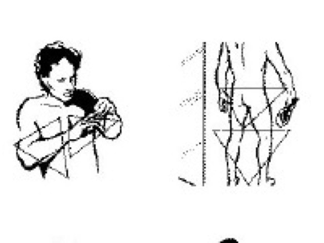
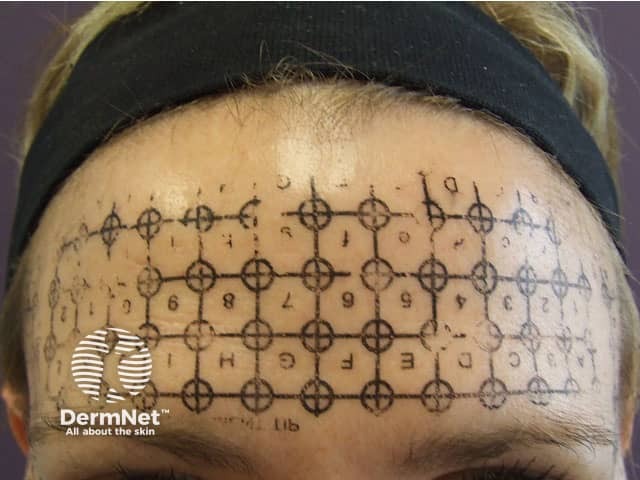
"Angry back" – multiple positive reactions due to eczema being too active
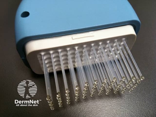
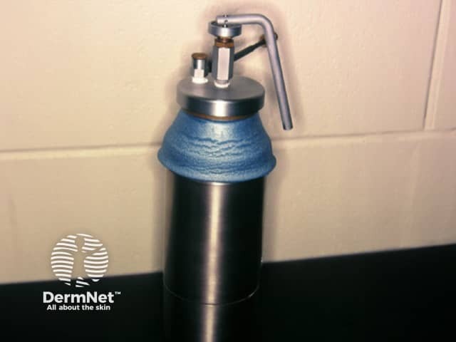
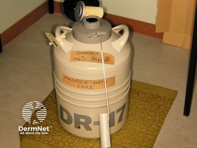
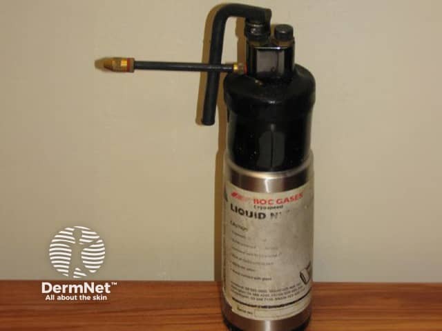
Cryotherapy liquid nitrogen dispenser
Close-up surgical shaving rash
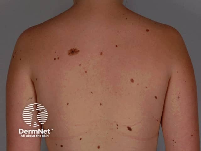
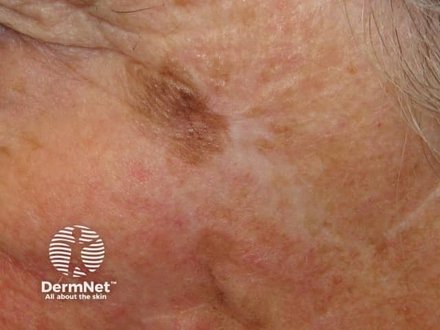
Recurring melanocytic naevus
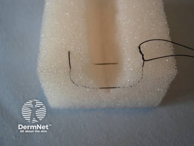
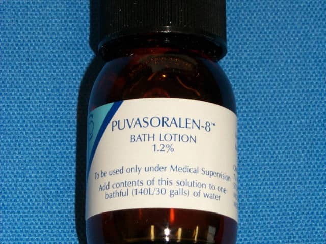
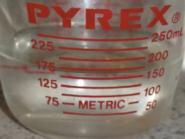
160 ml measured out for full bath
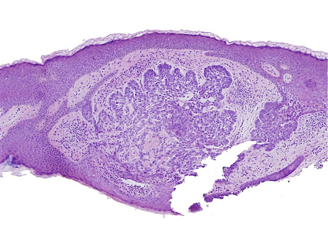
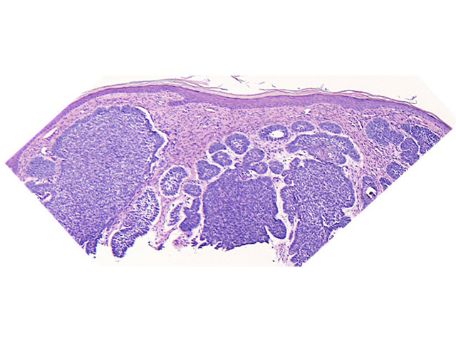
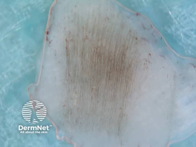
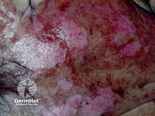
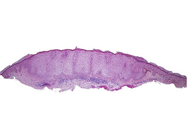
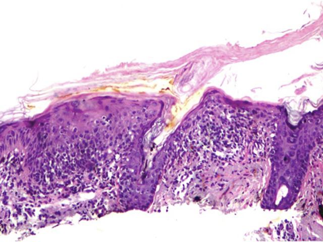
Histopathology of actinic keratosis
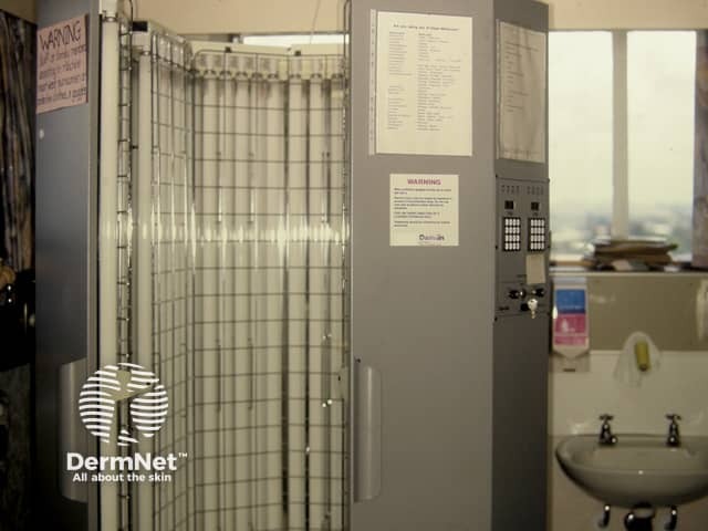
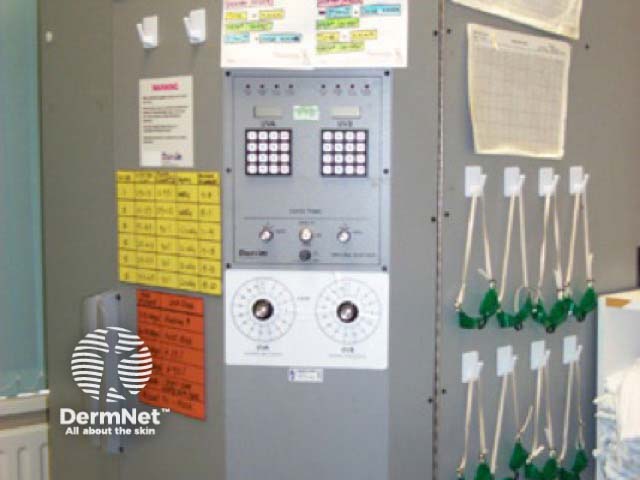
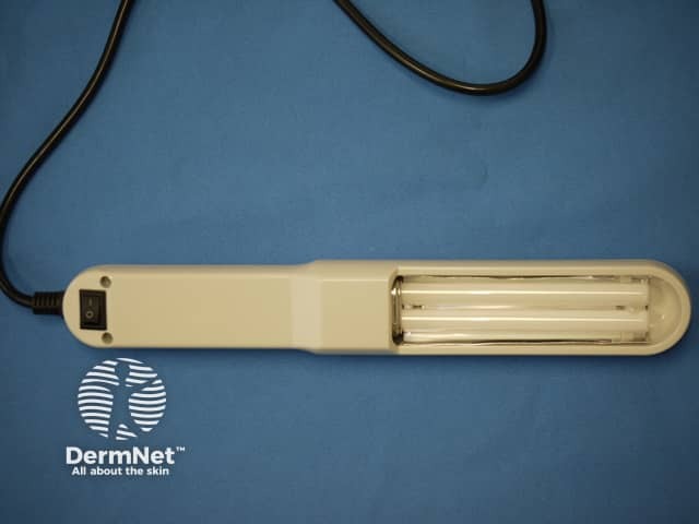
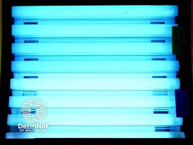
Unregulated Chinese narrowband UVB device
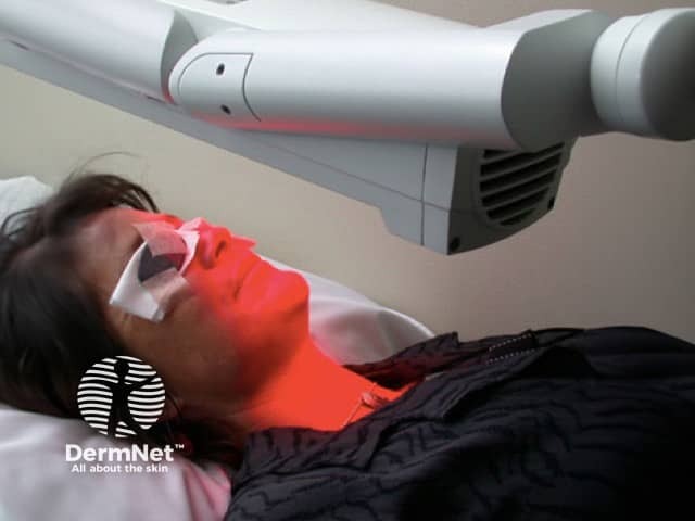
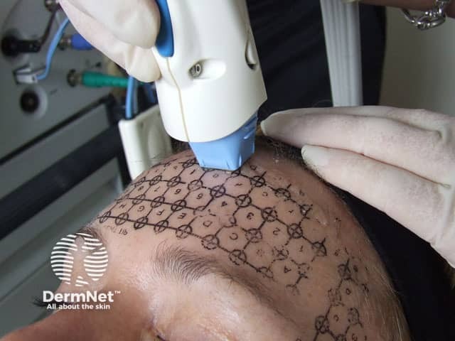
Radiation therapy being administed to a skin cancer on the arm
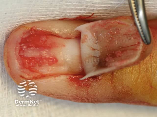
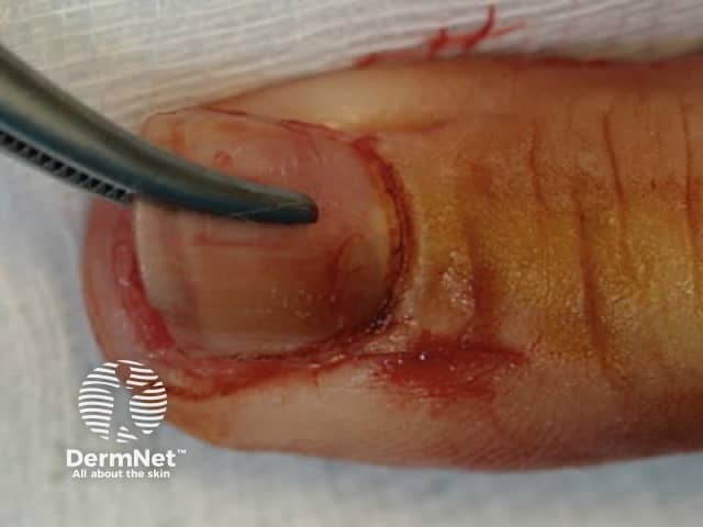
Hyperreflective patches, distal nail
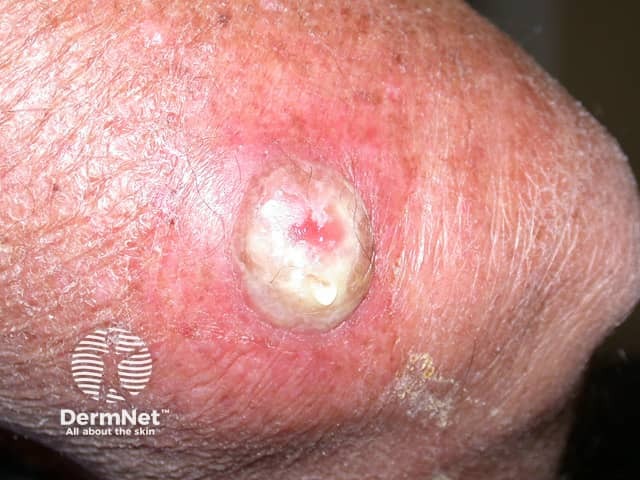
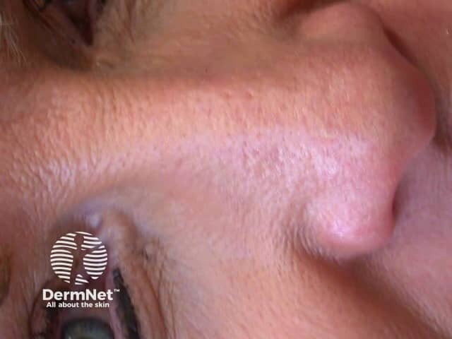
Superficial basal cell carcinoma
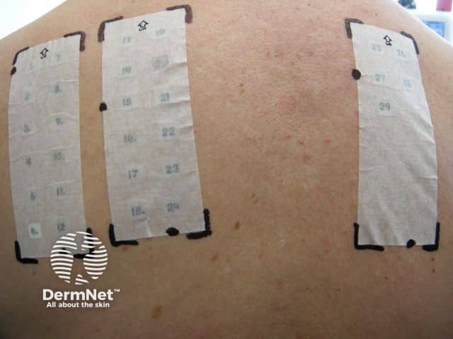
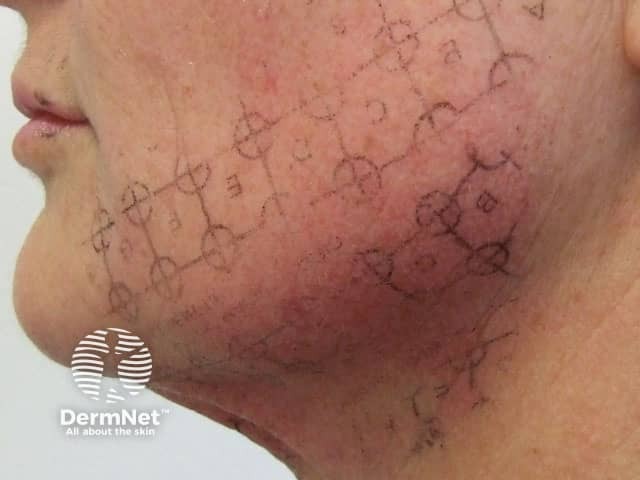
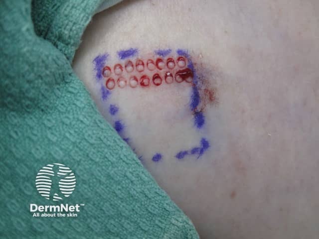
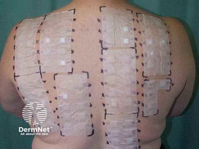
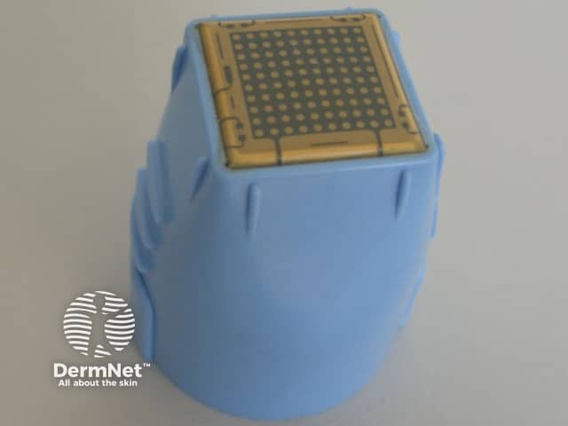
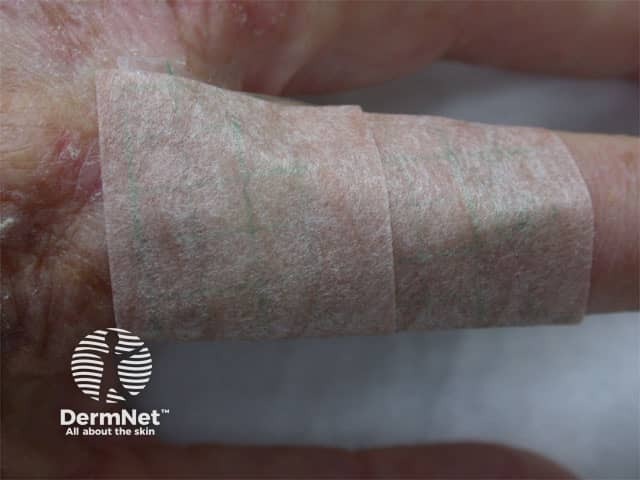
Patch test strips in place after application
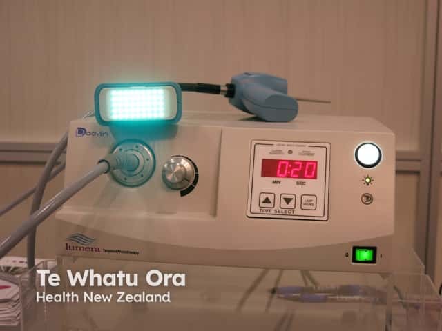
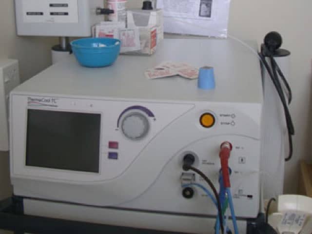
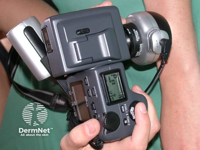
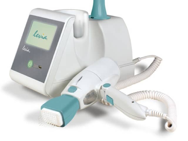
Pulsed dye laser treatment
Iontophoresis for palmar hyperhidrosis
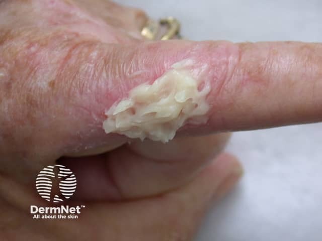
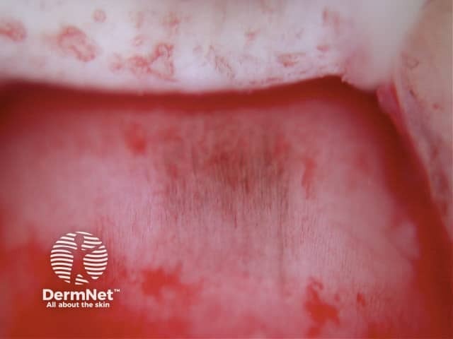
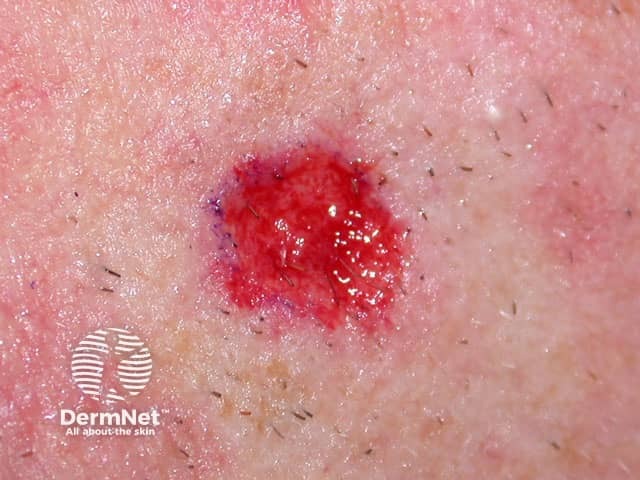
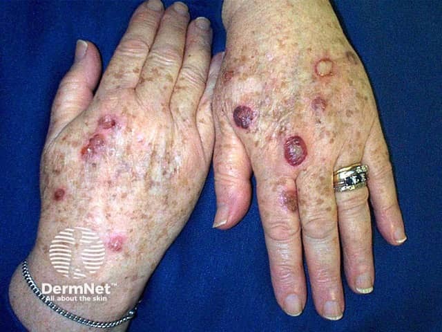
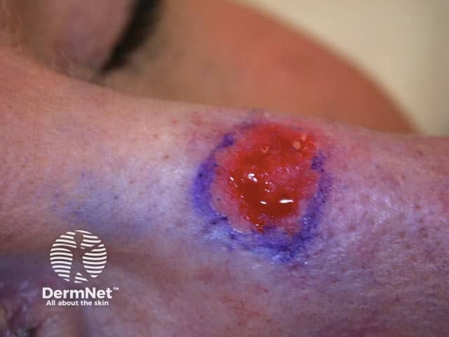
Haemorrhagic blisters 3 days after treatment
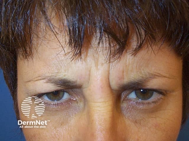
Subject frowning prior to botulinum toxin injectons
Pulse dye laser for telangiectasia, 2 weeks later
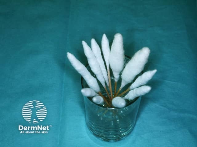
Large cotton swabs used for cryotherapy
Surgical wound dehiscence
Psoriasis disguised by cosmetic camouflage on right leg
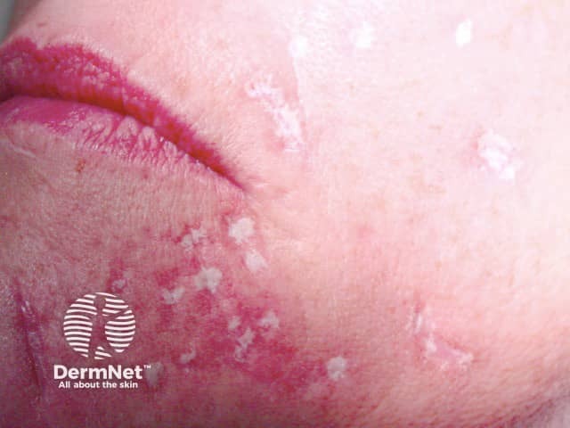
Frosting after applying trichloracetic acid in TCA CROSS procedure
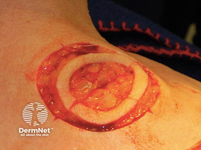
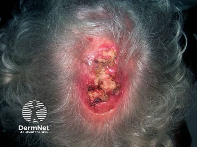
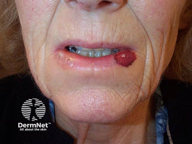
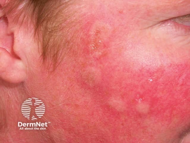
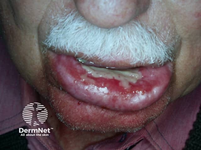
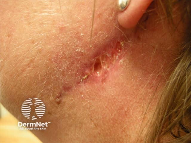
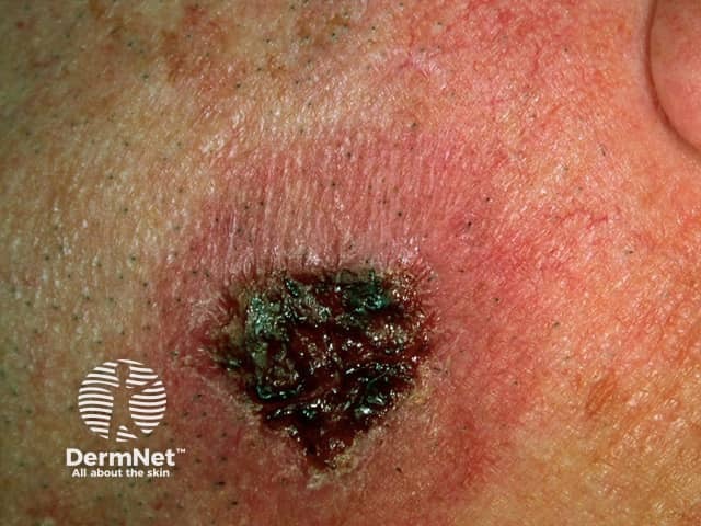
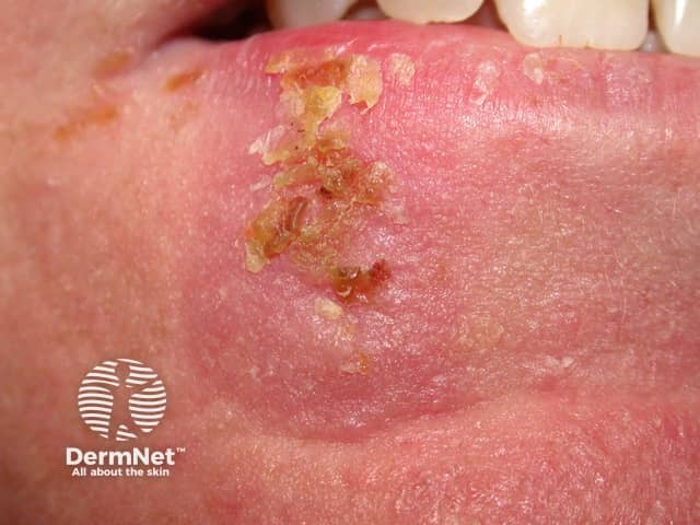
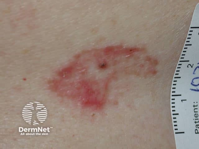
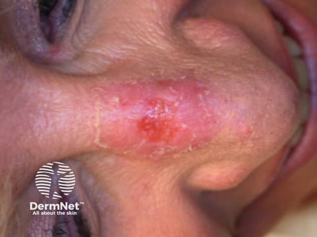
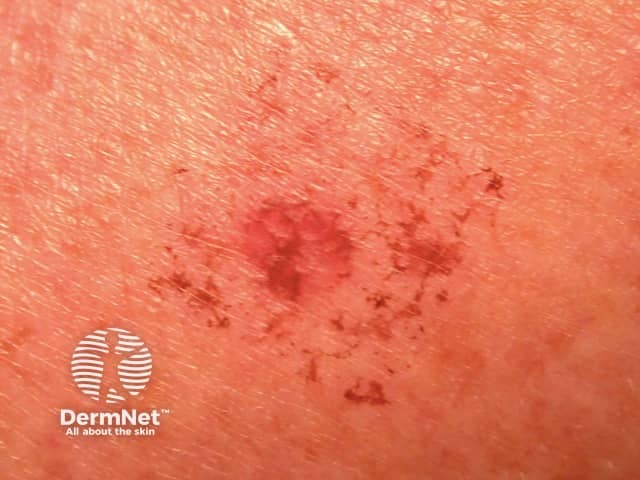
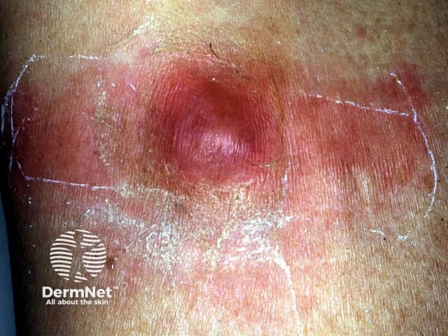
Abscess formed 2 months after
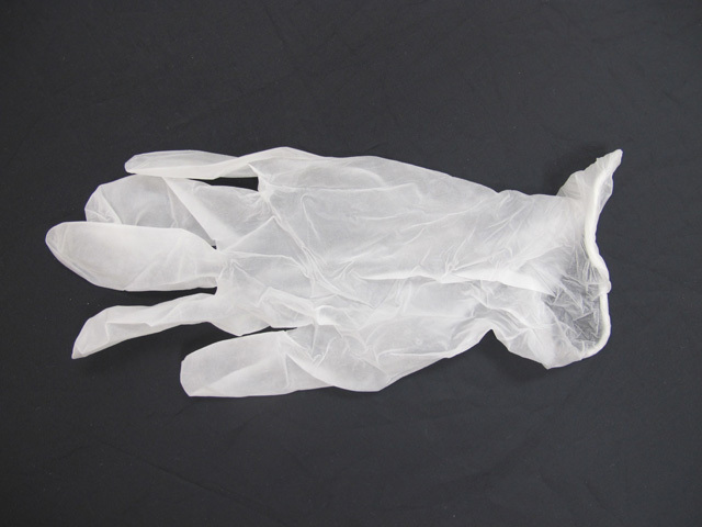
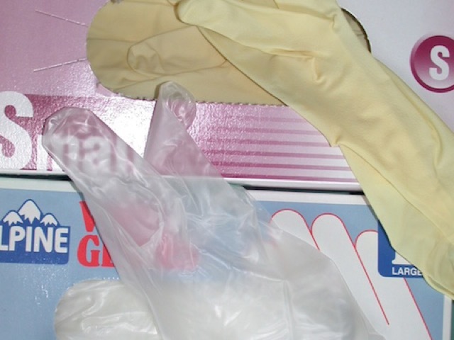
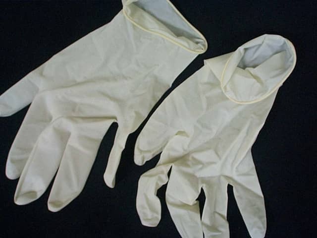
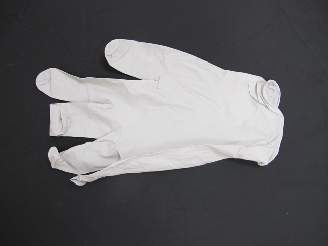
Nitrile examination gloves
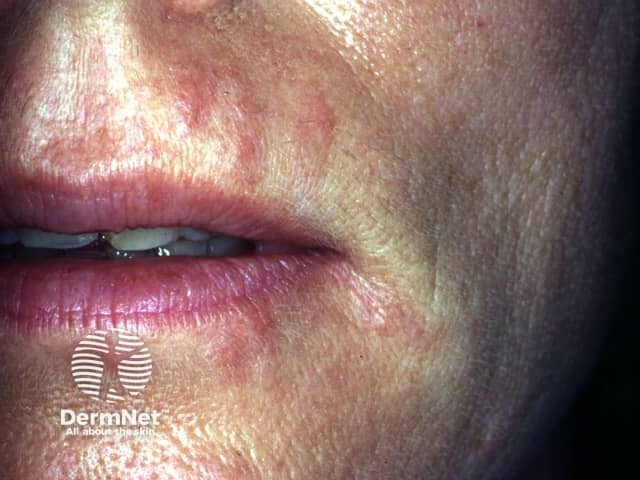
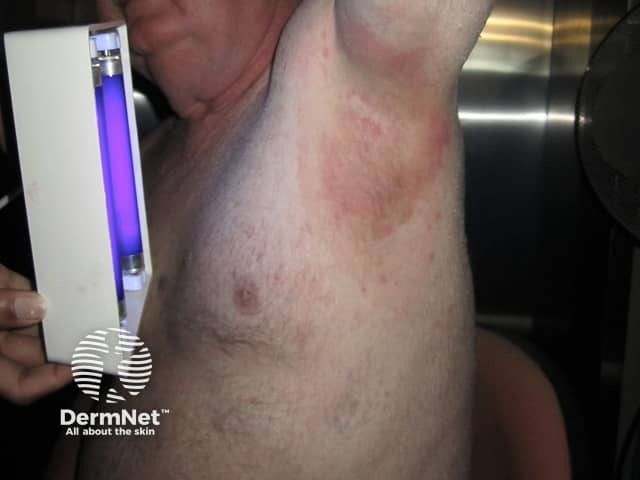
Coral-pink fluorescence in erythrasma using Wood lamp
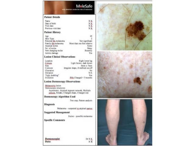
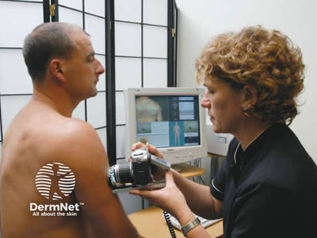
Macro and dermoscopy images of lesion of concern