Main menu
Common skin conditions

NEWS
Join DermNet PRO
Read more
Quick links

Figure 5
Keywords: Elastosis perforans serpiginosa, Histopathology-image, Pathology
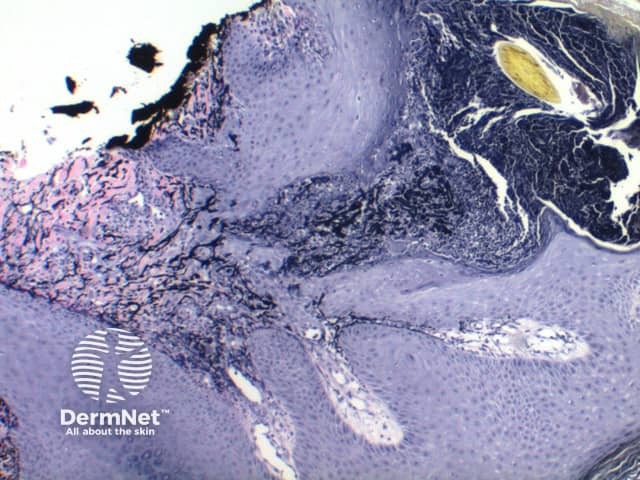
Low power of histology of elastosis perforans serpiginosa demonstrates a column of keratotic debris forming a focal invagination through a hyperplastic epidermis (figure 1). Closer inspection identifies material undergoing transepidermal elimination (figure 2). Brightly eosinophilic fibres are seen within the extruded material, mixed with keratinous debris and a mixed inflammatory cell infiltrate (figures 3, 4).
© DermNet
You can use or share this image if you comply with our image licence. Please provide a link back to this page.
For a high resolution, unwatermarked copy contact us here. Fees apply.
Source: dermnetnz.org