Figure 2
Keywords: Histopathology-image
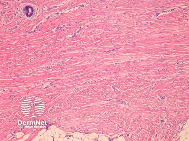
Fibroblastic connective tissue naevus is situated primarily in the reticular dermis, and often shows extension into the superficial subcutis (figures 1, 2). There may be a mild papillomatous epidermis. The lesional cells are bland spindle-shaped fibroblastic/myofibroblastic cells with indistinct pale eosinophilic cytoplasm and tapering nuclei, with no significant cytologic atypia or pleomorphism (figures 3, 4). These cells form short fascicles with no particular orientation to the overlying epidermis and are associated with a loose stroma composed of mainly delicate or wispy collagen bundles (figures 3, 4).
© DermNet
You can use or share this image if you comply with our image licence. Please provide a link back to this page.
For a high resolution, unwatermarked copy contact us here. Fees apply.
Source: dermnetnz.org

