

Syringofibroadenoma pathology
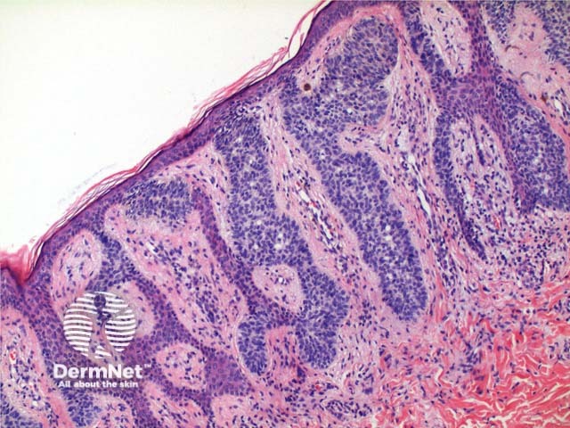
Figure 3
Keywords: Syringofibroadenoma, Histopathology-image, Pathology
Histology of syringofibroadenoma
Scanning power view of the histology of syringofibroadenoma reveals an epidermal proliferative process (Figure 1). Vertically oriented anastamosing strands of basaloid epithelium are seen arising from multiple points along the epidermis (Figure 2). The basaloid cells are smaller than adjacent keratinocytes and show mild variation in size (Figure 4). Ductal differentiation is seen (Figures 3 and 4). The tumour has notable intervening fibrovascular stroma, and may have a mild superficial lymphocytic infiltrate (Figure 4).
© DermNet
You can use or share this image if you comply with our image licence. Please provide a link back to this page.
For a high resolution, unwatermarked copy contact us here. Fees apply.
Source: dermnetnz.org