

Phaeohyphomycosis pathology
Figure 7
Keywords: Phaeohyphomycosis, Histopathology-image, Pathology
Histology of phaeohyphomycosis
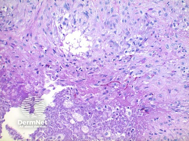
Scanning power view of phaeohyphomycosis shows a deeply extending granulomatous pattern (Figure 1) which may show areas of necrosis (Figure 2). Centrally an abscess or cystic nodule may form. Frequently a foreign body such as a wood splinter can be seen. The epidermis commonly shows pseudoepitheliomatous hyperplasia. The inflammatory infiltrate is comprised of histiocytes with multinucleated giant cells, and numerous neutrophils (Figures 3,4 and 5). At high power branching septate pigmented fungal hyphae can be seen (Figure 6).
© DermNet
You can use or share this image if you comply with our image licence. Please provide a link back to this page.
For a high resolution, unwatermarked copy contact us here. Fees apply.
Source: dermnetnz.org