

ADVERTISEMENT
Eccrine carcinoma pathology
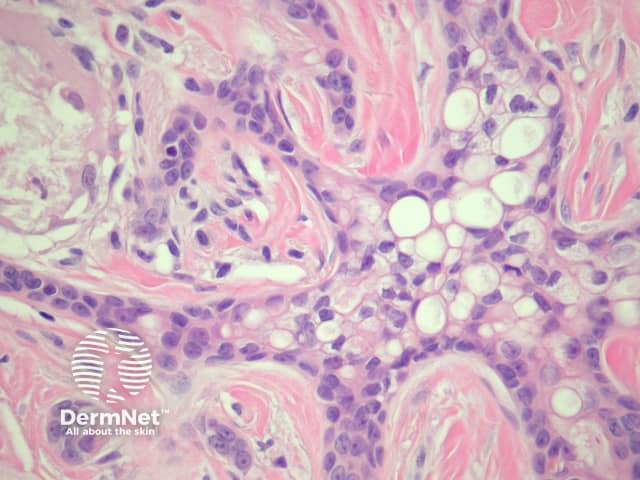
Figure 4
Keywords: Eccrine carcinoma, Histopathology-image, Pathology
Histology of eccrine carcinoma
Sections of eccrine carcinoma show a tumour composed of basaloid cells infiltrating the dermis (figure 1). The tumour forms tubules and glands (figures 2-4) which infiltrate a sclerotic dermis. Eccrine differentiation is readily seen at high power (best seen in figures 2 and 3). Clear cell change may be seen (figure 4).
© DermNet
You can use or share this image if you comply with our image licence. Please provide a link back to this page.
For a high resolution, unwatermarked copy contact us here. Fees apply.
Source: dermnetnz.org
ADVERTISEMENT