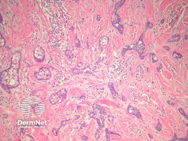
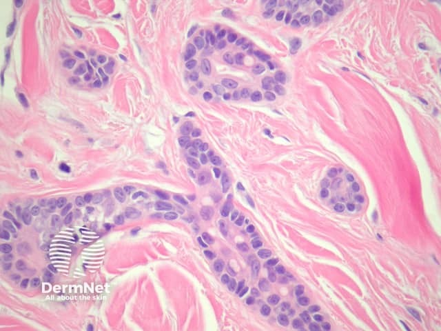
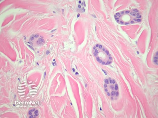
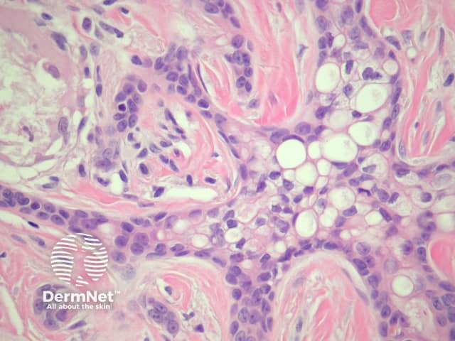
Sections of eccrine carcinoma show a tumour composed of basaloid cells infiltrating the dermis (figure 1). The tumour forms tubules and glands (figures 2-4) which infiltrate a sclerotic dermis. Eccrine differentiation is readily seen at high power (best seen in figures 2 and 3). Clear cell change may be seen (figure 4).
Immunohistochemical studies can be used to demonstrate glandular differentiation (CEA) in eccrine carcinoma, but are not needed if the morphology is classic. Some cases resemble breast cancer. Focal p63 and CK5/6 positivity is thought to favour a primary eccrine carcinoma.
Metastatic breast cancer – Clinical correlation may be needed to distinguish primary eccrine carcinoma from metastatic breast carcinoma. Immunohistochemical studies (see above) can be helpful
Microcystic adnexal carcinoma (MAC) – Some authors consider eccrine carcinoma to be a form of MAC. MAC generally demonstrates some squamous differentiation and keratinizing cystic changes superficially.
.

