Main menu
Common skin conditions

NEWS
Join DermNet PRO
Read more
Quick links

Trichoepithelioma pathology
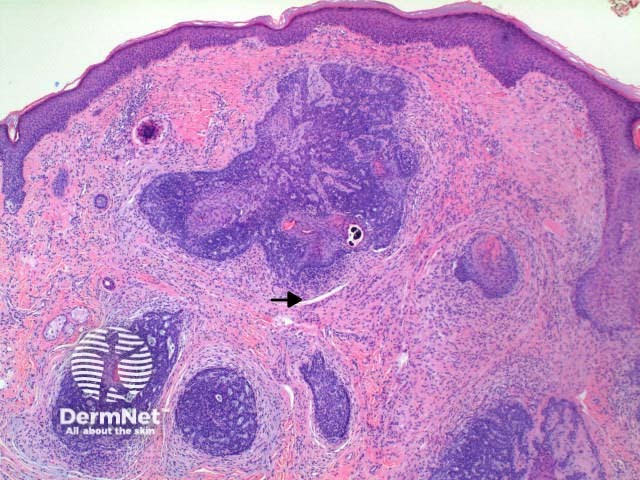
Figure 2
Keywords: Trichoepithelioma, Histopathology-image, Pathology
Histology of trichoepithelioma
Scanning power view reveals a tumour comprised of multiple nodules situated within the dermis (Figure 1). Small horn cysts, abortive hair follicles and calcification are frequently seen (Figure 2). The stroma is denser and more cellular than with basal cell carcinoma, and there is often focal stromal cracking (Figure 2, arrow). Often pronounced bulbar differentiation may be seen, emulating the follicular bulb and papilla; these structures have been referred to as papillary mesenchymal bodies. (Figure 3, arrow).
© DermNet
You can use or share this image if you comply with our image licence. Please provide a link back to this page.
For a high resolution, unwatermarked copy contact us here. Fees apply.
Source: dermnetnz.org
