Main menu
Common skin conditions

NEWS
Join DermNet PRO
Read more
Quick links

Porokeratosis pathology
Keywords: Porokeratosis, Histopathology-image, Pathology
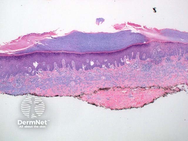
Scanning power view of porokeratosis reveals a hyperkeratotic lesion with a discrete parakeratotic column at the margin, or two if the whole lesion is represented (Figure 1). The diagnostic feature is the presence of a cornoid lamella which represents the clinically visible raised margin of the lesion. The cornoid lamella is a parakeratotic column overlying a small vertical zone of dyskeratotic and vacuolated cells within the epidermis (Figures 2 and 3). There is also a focal loss of the granular layer. A mild lymphocytic infiltrate may be seen around an increased number of capillaries in the underlying dermis.
© DermNet
You can use or share this image if you comply with our image licence. Please provide a link back to this page.
For a high resolution, unwatermarked copy contact us here. Fees apply.
Source: dermnetnz.org