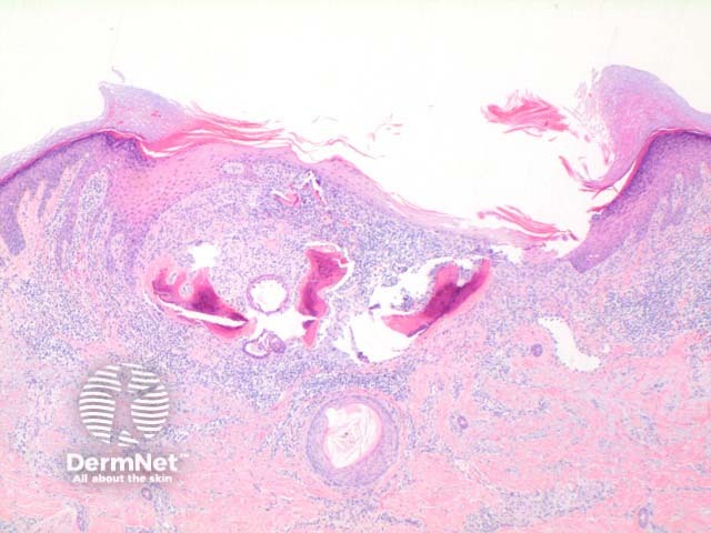
Figure 2
Keywords: Osteoma cutis, Histopathology-image, Pathology
Scanning power of osteoma cutis reveals the presence of dense eosinophilic deposits in the dermis or subcutaneous tissue (Figure 1). Spicules of bone may be seen to perforate the epidermis in the process of transepidermal elimination (Figures 2 and 3). Most cutaneous bone formation occurs by the process of membranous ossification and so associated cartilage tissue is lacking. Bone is identified by osteocytes held within small lacunae (Figures 4 and 5) and the hydroxyapatite eosinophilic support material. In larger deposits, Haversian systems can be seen as concentric osteocytes around a central blood vessel (carried within a Haversian canal).
© DermNet
You can use or share this image if you comply with our image licence. Please provide a link back to this page.
For a high resolution, unwatermarked copy contact us here. Fees apply.
Source: dermnetnz.org

