Main menu
Common skin conditions

NEWS
Join DermNet PRO
Read more
Quick links

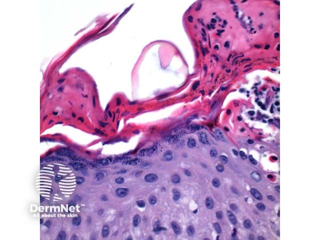
Figure 4
Keywords: Histopathology-image, Pathology, Scabies
Scanning power view of scabies shows a pattern of an epidermal and wedge shaped dermal inflammatory process (Figure 1). The epidermis may show significant scale crust comprised of serous exudate, neutrophils, and eosinophils (Figure 2). There may be focal ulceration or erosion secondary to excoriation. The inflammatory infiltrate may show a wedge shaped or diffuse superficial and deep perivascular and interstitial pattern. Lymphocytes with numerous eosinophils are the rule with scattered superficial neutrophils seen in excoriated or impetiginised cases. Deep interstitial eosinophils are an important clue to an arthropod bite reaction (Figure 3).
© DermNet
You can use or share this image if you comply with our image licence. Please provide a link back to this page.
For a high resolution, unwatermarked copy contact us here. Fees apply.
Source: dermnetnz.org