Figure 1
Keywords: Trichilemmal carcinoma, Histopathology-image, Pathology
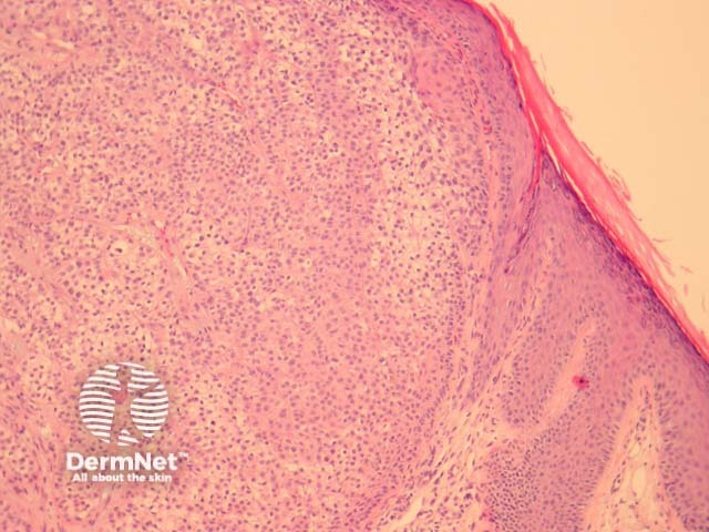
Trichilemmal carcinoma is often well defined laterally and is composed of lobules of atypical keratinocytes exhibiting clear cell change. At the periphery of the lobules the keratinocytes show palisading and are surrounded by a prominent connective tissue sheath (figures 1, 2). The invasive tumour front is usually a broad pushing front. The cells often exhibit marked pleomorphism and numerous mitoses (figure 3, 4).
© DermNet
You can use or share this image if you comply with our image licence. Please provide a link back to this page.
For a high resolution, unwatermarked copy contact us here. Fees apply.
Source: dermnetnz.org

