Main menu
Common skin conditions

NEWS
Join DermNet PRO
Read more
Quick links

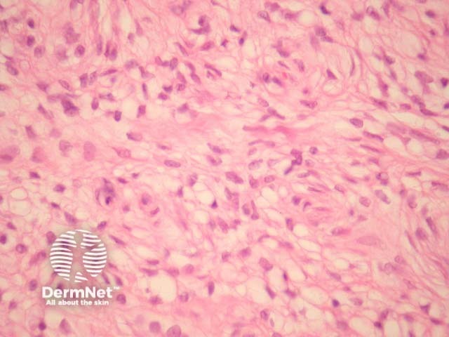
Figure 2
Keywords: Perineurioma, Histopathology-image, Pathology
Perineurioma tumour cells are spindle shaped, with long delicate cell processes amidst a collagenous stroma (figure 1). The spindle cells have pale open nuclei with a delicate chromatin pattern, an inconspicuous eosinophilic nucleolus and indistinct cell borders (figures 1, 2). Myxoid change may be seen and can cause diagnostic confusion (figure 3). Whorls of cells are often seen which are reminiscent of meningioma (figure 4, arrows).
© DermNet
You can use or share this image if you comply with our image licence. Please provide a link back to this page.
For a high resolution, unwatermarked copy contact us here. Fees apply.
Source: dermnetnz.org