Main menu
Common skin conditions

NEWS
Join DermNet PRO
Read more
Quick links

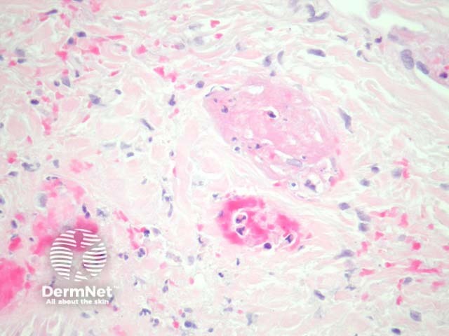
Figure 2
Keywords: Warfarin necrosis, Histopathology-image, Pathology
In warfarin necrosis, sections show variable degrees of epidermal and dermal necrosis (figure 1). There are extensive intravascular thrombi within capillaries and venules (figures 2, 3). There remaining patent vessels are dilated.
© DermNet
You can use or share this image if you comply with our image licence. Please provide a link back to this page.
For a high resolution, unwatermarked copy contact us here. Fees apply.
Source: dermnetnz.org