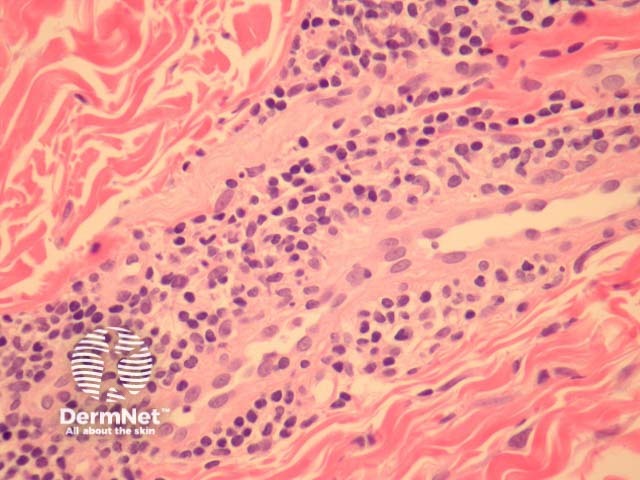
Figure 2
Keywords: Perniosis, Histopathology-image, Pathology
In perniosis, sections show acral skin with a dense superficial and deep lymphocytic infiltrate (figure 1). Subepidermal oedema may be marked. The characteristic feature is lymphocytic perivascular infiltrate within the dermis and sometimes extending to the subcutis (figures 2, 3). This is thought by some authors to represent the one true “lymphocytic vasculitis”, as there may be well established fibrinoid change and thrombosis can occur. There may be numerous eosinophils in early lesions (figure 4).
© DermNet
You can use or share this image if you comply with our image licence. Please provide a link back to this page.
For a high resolution, unwatermarked copy contact us here. Fees apply.
Source: dermnetnz.org

