Figure 4
Keywords: Non-clinical-image
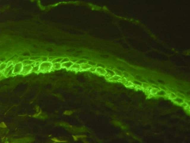
Early lesions of pemphigus vulgaris show suprabasal epidermal acantholysis, clefting and blister formation. The blister cavity may contain inflammatory cells including eosinophils and rounded acantholytic cells with intensely eosinophilic cytoplasm and a perinuclear halo. The floor of the blister may be lined with intact keratinocytes, the “tombstone pattern” (figures 1, 2). Acantholysis can also affect adnexae. Dermal changes include perivascular inflammatory infiltrate particularly with eosinophils.
Open access image
You can use or share this image if you comply with our image licence. Please provide a link back to this page.
For a high resolution, unwatermarked copy contact us here. Fees apply.

